.webp&w=384&q=75)

Loading, please wait...
Loading, please wait...

Idiopathic pulmonary fibrosis remains a devastating interstitial lung disease characterized by progressive extracellular matrix deposition and respiratory failure. In healthy alveolar spaces, abundant oxygen normally triggers rapid degradation of hypoxia-inducible factor 1-alpha. However, recent evidence reveals that alveolar macrophages in fibrotic lungs continuously stabilize this factor despite existing within a well-oxygenated environment. This physiological contradiction highlights an anomalous pseudohypoxic state driven by environmental and metabolic triggers. In particular, ROS-HIF-1 signalling plays a critical role in converting oxidative stress into sustained transcriptional changes. Reactive oxygen species disrupt normal oxygen-sensing machinery, forcing resident immune cells to adopt pathogenic phenotypes. Consequently, these rewired macrophages impair tissue repair and actively sustain chronic inflammation. Clinicians must recognize how inhaled environmental toxins and endogenous metabolic disturbances perpetuate this pathway. Understanding this intracellular circuit provides crucial insights into how early epithelial and macrophage injuries culminate in terminal pulmonary architectural remodeling.
Under baseline conditions, prolyl hydroxylase domain enzymes utilize molecular oxygen and ferrous iron to hydroxylate key proline residues on hypoxia-inducible factor subunits. Subsequently, the Von Hippel-Lindau protein targets hydroxylated factor subunits for proteasomal destruction. In idiopathic pulmonary fibrosis, however, this regulatory balance fails completely within alveolar macrophages. Excessive generation of reactive oxygen species directly oxidizes the ferrous iron cofactor required by prolyl hydroxylases. As a result, hydroxylase catalytic activity declines, which prevents ubiquitination and degradation of the master transcription factor. Therefore, macrophages experience functional hypoxia while bathed in atmospheric alveolar oxygen tensions. This pseudohypoxic state decouples macrophage genetic responses from true tissue oxygen availability. Moreover, inhaled particulates, environmental pollutants, and cigarette smoke amplify this oxidative burden significantly. By shifting cellular perception away from ambient oxygen levels, oxidative stress sustains a perpetual survival and activation program that compromises normal lung homeostasis.
When ROS-HIF-1 signalling remains chronically active, alveolar macrophages undergo profound metabolic and immunological reprogramming. Normally, tissue-resident alveolar macrophages rely primarily on mitochondrial oxidative phosphorylation to perform surfactant clearance and anti-inflammatory clearance duties. In contrast, stabilized transcription factors induce a pronounced glycolytic shift. This metabolic switch markedly accelerates the transcription of proinflammatory and profibrotic cytokines. Specifically, reprogrammed macrophages release excessive amounts of transforming growth factor-beta, platelet-derived growth factor, and plasminogen activator inhibitor-1 into the interstitium. Consequently, adjacent lung fibroblasts proliferate rapidly and differentiate into collagen-secreting myofibroblasts. In addition, the loss of normal homeostatic phagocytosis prevents effective clearance of apoptotic debris and epithelial fragments. Thus, the macrophage transitions from a protective sentinel into an aggressive driver of fibrogenesis. This continuous paracrine crosstalk relentlessly stiffens the parenchymal matrix and erodes gas exchange capacity across the alveolar membrane.
The molecular regulation of prolyl hydroxylase activity depends strictly upon intracellular redox balance and iron homeostasis. Intracellular reactive oxygen species, such as hydrogen peroxide and superoxide anions, convert catalytic ferrous iron into an inactive ferric state. Consequently, the hydroxylase active site loses its ability to transfer oxygen atoms onto target proline residues. Furthermore, dysregulated cellular iron handling frequently accompanies chronic lung disease and promotes the Fenton reaction. This chemical cascade generates highly reactive hydroxyl radicals that further damage cellular membranes and inactivate metabolic enzymes. In addition, endogenous antioxidants like glutathione become rapidly depleted during continuous oxidative challenges. Therefore, endogenous antioxidant defenses cannot neutralize the sustained radical production observed in fibrotic tissue niches. Because hydroxylase suppression becomes irreversible under profound oxidative stress, transcriptional activation persists unabated. Targeting these precise enzymatic and iron-dependent nodes offers an attractive framework for novel therapeutic strategies aimed at halting relentless lung scar formation.
Current antifibrotic treatments, including pirfenidone and nintedanib, slow disease progression but fail to reverse established fibrosis or halt mortality entirely. Consequently, exploring non-classical upstream drivers of macrophage activation has become a primary translational priority. Interventions that target the intersection of oxidative stress and transcriptional stabilization hold tremendous promise for future clinical management. Specifically, cell-permeable antioxidants and targeted mitochondrial scavengers can reduce cytoplasmic radical accumulation in alveolar macrophages. Furthermore, agents that restore ferrous iron pools or directly enhance hydroxylase activity may restore normal transcription factor turnover. Inhaled drug delivery formulations represent an especially appealing strategy because they deposit high local therapeutic concentrations directly into alveolar airspaces. By neutralizing the initial trigger within resident immune cells, clinicians could theoretically arrest fibrotic cascades before irreversible architectural remodeling occurs. Continued interdisciplinary research across cellular biology and respiratory pharmacology will clarify whether disrupting these pathways can improve long-term clinical survival.
Pseudohypoxia occurs when alveolar macrophages exhibit hypoxia-inducible factor stabilization despite adequate ambient oxygen concentrations. Reactive oxygen species oxidize catalytic ferrous iron within prolyl hydroxylase enzymes, preventing standard factor degradation. Consequently, the macrophage behaves metabolically as though oxygen were depleted. This dysregulation initiates harmful downstream inflammatory cascades, stimulates excessive extracellular matrix production, and accelerates progressive fibrotic remodeling throughout the surrounding lung architecture.
Excessive reactive oxygen species drive a metabolic shift known as the Warburg effect by stabilizing transcription factors. Rather than utilizing quiescent mitochondrial oxidative phosphorylation, alveolar macrophages switch predominantly to aerobic glycolysis. Furthermore, this metabolic reconfiguration alters cellular cytokine production, markedly upregulates profibrotic mediators like transforming growth factor-beta, and reduces the natural capacity of the macrophage to clear surfactant and apoptotic debris.
Targeting macrophage signalling offers significant therapeutic potential alongside current standard antifibrotic pharmacotherapies. By utilizing targeted radical scavengers or restoring prolyl hydroxylase enzyme function, clinicians could prevent the abnormal phenotypic reprogramming of resident alveolar immune cells. Consequently, disrupting this circuit may diminish the paracrine stimulation of pulmonary fibroblasts, decrease progressive lung stiffness, and ultimately help preserve vital gas exchange capacity in patients.
Disclaimer: This content is for informational and educational purposes only. It is not intended to substitute for professional medical advice, diagnosis, or treatment. Always seek the advice of your physician or another qualified healthcare provider with any questions you may have regarding a medical condition or treatment options. Never disregard professional medical advice or delay in seeking it because of something you have read here. Refer to the latest local and national guidelines for clinical practice.
References
E J et al. ROS/HIF-1 signalling in alveolar macrophages: implications for lung fibrosis. Am J Physiol Lung Cell Mol Physiol. 2026 Sep 24. doi: 10.1152/ajplung.00192.2026. PMID: 42779491.
Adán-Barrientos I, et al. HIF-1α and HIF-2α transcription factors differentially regulate lung alveolar macrophage function. J Clin Invest. 2024;134(8):e171802.
Wang Y, et al. Hypoxia-inducible factor signaling in inflammatory lung injury and repair. Antioxidants (Basel). 2022;11(1):114.

Read summarized clinical updates, watch expert medical content, and earn CME certifications right from your smartphone.
Open your smartphone camera to scan and install.





Alveolar macrophages in idiopathic pulmonary fibrosis display aberrant HIF-1α stabilization despite residing in oxygen-rich alveoli. Driven by reactive oxygen species, this pseudohypoxic signalling reprograms macrophages toward a profibrotic phenotype, creating new translational avenues for pulmonary therapy.
Today

The ClinGen Prenatal Gene Curation Expert Panel evaluated 63 disease relationships across 61 genes, establishing clinical validity for severe fetal phenotypes like hydrops and stillbirth to enhance prenatal genomic interpretation and clinical care.
Today

A cohort study demonstrates that the weight-adjusted waist index predicts all-cause and cardiovascular mortality more accurately than BMI and conventional adiposity metrics, offering clinicians a practical anthropometric tool for superior cardiometabolic risk stratification.
Today

New research reveals that HOXD13 regulates primary cilia signalling and genomic stability in distal synovial fibroblasts, uncovering why conditions like rheumatoid arthritis target specific joints.
Today
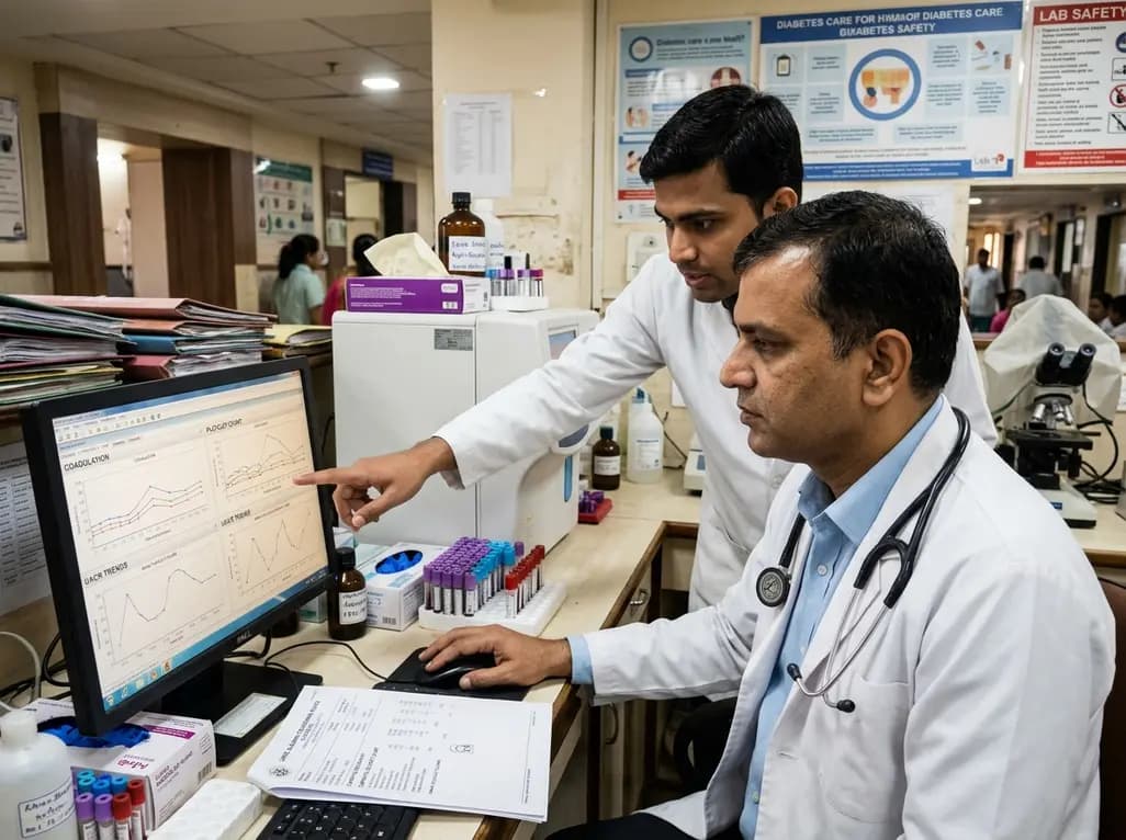
A western India study demonstrates that shortened coagulation times (PT, APTT), elevated mean platelet volume, and increased UACR strongly correlate with type 2 diabetes microvascular complications, highlighting the clinical value of combined hemostatic and renal profiling for early risk stratification.
Today
