

Loading, please wait...
Loading, please wait...

A right aortic arch variant represents an uncommon congenital vascular anomaly that clinicians usually discover incidentally during adulthood. While normal embryogenesis produces a left-sided aortic arch with three primary branches, anatomical anomalies occur when primitive branchial structures regress abnormally. In a recently reported case, clinicians identified a distinctive vascular configuration in a 48-year-old female presenting for acute neurovascular stroke assessment. Arch-to-vertex computed tomography angiography established three consecutive branches arising directly from the right aortic arch. First, a true bicarotid trunk emerged to bifurcate cleanly into both common carotid arteries. Second, a separate right subclavian artery originated from the arch. Third, an aberrant left subclavian artery arose distally and pursued a retroesophageal course toward the left hemithorax. Importantly, thorough vascular evaluation confirmed the absence of aortic dissection or an associated Kommerell diverticulum. Although acute stroke prompted radiological imaging, neurovascular teams found no direct causal link between the arch architecture and the cerebrovascular event. Instead, this presentation underscores how advanced imaging protocols clarify complex supra-aortic branching patterns in symptomatic adult patients.
Understanding embryological vascular remodeling clarifies why this unusual anatomy develops during early gestation. During normal development, six pairs of branchial arches and two dorsal aortae undergo coordinated regression and persistence. A right-sided arch typically develops when the right fourth branchial arch persists while the left fourth arch regresses prematurely. In this specific configuration, clinicians observe two distinct embryological phenomena. First, the ventral aortic roots coalesce unusually, creating a true bicarotid trunk that gives rise to both common carotid arteries without incorporating subclavian vessels. Consequently, this trunk differs fundamentally from a bovine arch, which features a common origin of the brachiocephalic artery and left common carotid artery. Second, regression of the left fourth arch forces the left subclavian artery to originate abnormally from the proximal descending dorsal aorta. Therefore, the vessel must navigate across the posterior mediastinum toward the left arm. Because this retroesophageal route traverses confined thoracic compartments, it can compress adjacent mediastinal viscera under specific hemodynamic or mechanical conditions.
Modern cross-sectional imaging provides definitive anatomical clarity when evaluating complex thoracic vascular patterns. While conventional axial imaging offers basic structural cross-sections, it often fails to resolve intricate vessel takeoffs and tortuous mediastinal trajectories. Therefore, arch-to-vertex computed tomography angiography plays an indispensable diagnostic role. Using thin-slice volumetric acquisition, multiplanar reformations, and three-dimensional volume-rendered reconstructions, radiologists meticulously map the precise origin and path of each branch. In this case, 3D reconstructions definitively distinguished a true bicarotid trunk from other common supra-aortic variants. Furthermore, high-resolution imaging permitted precise exclusion of a Kommerell diverticulum, which often forms an aneurysmal outpouching at the origin of an aberrant subclavian vessel. In addition, contrast-enhanced computed tomography angiography swiftly ruled out concurrent aortic dissection and mural thrombus. Consequently, radiologists and neurovascular teams could ascertain the patient's vascular geometry with absolute fidelity. This rapid, non-invasive imaging approach eliminates diagnostic ambiguity and ensures optimal guidance for both emergency diagnostics and elective interventional pathways.
An aberrant subclavian artery often remains clinically silent throughout childhood, but it can trigger symptoms later in adult life. As patients age, progressive arterial stiffening, atherosclerosis, or secondary aortic tortuosity can accentuate mechanical pressure against neighboring structures. In this patient, focused clinical questioning disclosed a chronic history of swallowing difficulties, commonly designated as dysphagia lusoria. However, detailed imaging failed to demonstrate definitive, fixed extrinsic compression of the esophageal lumen. Clinicians must recognize that intermittent functional compression during bolus transit can induce symptoms without showing severe static luminal collapse on supine imaging. Meanwhile, the patient's acute stroke required rigorous investigation to exclude potential hemodynamic or embolic causes linked to the aberrant vessels. Although severe carotid tortuosity can occasionally promote local turbulent flow, diagnostic workup established no causal relationship with the ischemic event. Therefore, clinicians must avoid prematurely attributing neurological deficits to incidental congenital variations. Instead, they should maintain a disciplined diagnostic framework that thoroughly evaluates classical cardiovascular risk factors alongside rare structural anomalies.
Accurate identification of a right aortic arch variant holds profound practical significance for cardiovascular, thoracic, and neurointerventional specialists. When interventionalists perform emergency mechanical thrombectomy or elective carotid stenting, atypical arch branches pose substantial navigation hurdles. Specifically, standard transfemoral catheter trajectories often encounter acute vessel angulations at the ostium of a bicarotid trunk. Consequently, endovascular operators may need to select specialized guiding catheters, modified microwires, or alternative radial and brachial access sites. Similarly, cardiac and vascular surgeons must anticipate this aberrant layout prior to performing mediastinal operations, tracheal resections, or aortic repairs. Unrecognized retroesophageal vessels face an elevated risk of accidental transection, acute ischemia, or massive intraoperative hemorrhage. Moreover, anesthesiologists and critical care physicians must exercise caution during transesophageal echocardiography, nasogastric tube placement, and central venous catheterization. By clearly identifying these variations beforehand through advanced computed tomography angiography, multidisciplinary hospital teams minimize procedural delays, reduce intraoperative complications, and optimize individualized patient safety across complex clinical scenarios.
A true bicarotid trunk represents an isolated aortic branch that divides exclusively into both the right and left common carotid arteries. In contrast, the common anatomical pattern known as a bovine arch typically involves a shared origin of the brachiocephalic trunk and the left common carotid artery. Understanding this fundamental anatomical distinction helps interventional radiologists select appropriate catheter shapes during transfemoral endovascular procedures.
An aberrant left subclavian artery arises from the distal arch and crosses the posterior mediastinum to reach the left upper extremity. Because the artery passes behind the esophagus, progressive age-related vascular stiffness or atherosclerotic expansion can compress the flexible esophageal wall. This mechanical impaction impedes food bolus transit, resulting in the classic chronic swallowing difficulties termed dysphagia lusoria.
Computed tomography angiography provides high-resolution, multiplanar, and three-dimensional volumetric images of thoracic and cervical vasculature. These detailed projections allow clinicians to accurately map aberrant vessel origins, evaluate retroesophageal trajectories, and inspect luminal patency. Furthermore, CT angiography reliably rules out critical vascular complications such as Kommerell diverticulum, aneurysm formation, or aortic dissection prior to planning surgical or endovascular interventions.
Disclaimer: This content is for informational and educational purposes only and is not intended to serve as medical advice, diagnosis, or treatment. Healthcare professionals should rely on their clinical judgment and verify details against relevant pharmacological and institutional protocols. Treatment decisions must always be tailored to individual patient needs. The views expressed here do not necessarily reflect the official policies of any medical board or governing body. Refer to the latest local and national guidelines for clinical practice.
References

Read summarized clinical updates, watch expert medical content, and earn CME certifications right from your smartphone.
Open your smartphone camera to scan and install.





Arch-to-vertex CT angiography revealed a rare right aortic arch variant with a true bicarotid trunk and aberrant left subclavian artery in an adult with stroke and chronic dysphagia. The case highlights crucial diagnostic and procedural implications for vascular, surgical, and neurointerventional teams.
Today
Metal-dependent cell death modalities like ferroptosis and cuproptosis dictate immune recognition in cancer. By integrating cell-derived danger signals and local metal dynamics, these pathways can transform immune-cold lesions into responsive tumors, providing new therapeutic avenues to overcome immunotherapy resistance.
Today

A prospective study reveals that over 54% of women with non-celiac wheat sensitivity experience uro-gynecological disorders, including dysmenorrhea and abacterial cystitis. Adherence to a wheat-free diet provides significant, long-term symptom resolution over 24 months.
Today
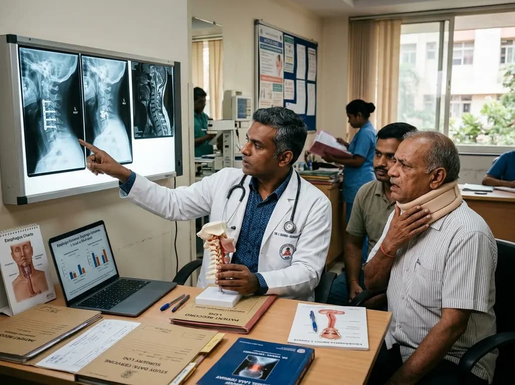
A large registry study reveals that postoperative dysphagia occurs more frequently and persists longer after multilevel anterior cervical spine surgery than single-level procedures. Key risk factors include baseline dysphagia, diabetes, upper cervical segment involvement, and medical comorbidities.
Today
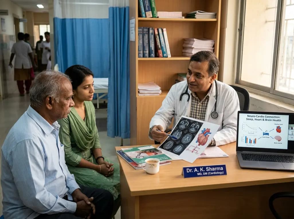
A landmark UK Biobank study by Karolinska Institutet researchers reveals that optimal rest mitigates neurodegeneration in cardiometabolic illness. Patients maintaining high sleep quality demonstrated superior cognitive scores and larger brain volumes, highlighting sleep optimization as an actionable clinical strategy.
Today
