.webp&w=384&q=75)

Loading, please wait...
Loading, please wait...

Applying radiomics lower grade glioma workflows provides powerful prognostic insights for patients with isocitrate dehydrogenase (IDH) mutations. Adult diffuse lower-grade gliomas present significant clinical heterogeneity, which routinely complicates postoperative management. Consequently, neuro-oncologists and neurosurgeons face a delicate clinical balancing act. Treating all patients immediately with adjuvant radiation and chemotherapy increases the risk of severe long-term neurocognitive toxicity. Conversely, delaying postoperative intervention may compromise overall survival when aggressive tumor biology remains undetected. Therefore, clinicians urgently require refined prognostic instruments to calibrate treatment timing effectively. Conventional magnetic resonance imaging offers essential baseline anatomical details. However, standard visual inspection fails to capture subtle subvisual variations in tumor architecture. In contrast, advanced computational extraction translates routinely acquired radiological images into objective, quantifiable data matrices. By revealing hidden microstructural patterns, machine learning algorithms empower clinicians to anticipate clinical trajectories with greater confidence. As a result, radiomics serves as a crucial bridge between structural imaging and precision neuro-oncology. Furthermore, recent institutional investigations emphasize that non-invasive phenotypic characterization can substantially improve risk stratification. Multidisciplinary neuro-oncology tumor boards will benefit substantially from objective algorithms that reliably differentiate indolent presentations from high-risk neoplasms before initiating toxic adjuvant regimens.
Diffuse lower-grade gliomas carrying IDH mutations encompass astrocytomas and oligodendrogliomas with remarkably diverse disease courses. Although these neoplasms frequently manifest in young, active adults with preserved performance status, malignant transformation remains an inevitable threat. Consequently, clinicians constantly debate the ideal therapeutic window for postoperative management. Immediate administration of radiotherapy and alkylating chemotherapy effectively suppresses recurrence. However, these aggressive interventions frequently cause irreversible white matter damage, secondary malignancies, and severe neurocognitive decline. Conversely, a watchful waiting approach preserves cognitive functions initially, but it exposes certain patients to untimely tumor progression. Thus, traditional prognostic variables often fail to resolve this therapeutic uncertainty. Standard prognostic assessments rely heavily on patient age, baseline functional score, histological grade, and gross total resection. Nevertheless, these clinical variables do not fully expose underlying intratumoral biological heterogeneity. In addition, two tumors with identical macroscopic appearances can follow divergent clinical trajectories over five to ten years. Therefore, neuro-oncologists require quantitative, tumor-intrinsic biomarkers to distinguish aggressive phenotypes from indolent tumors accurately. Addressing this fundamental diagnostic gap will allow multidisciplinary teams to deliver timely therapy while avoiding premature toxicity in low-risk individuals.
To improve risk discrimination, investigators designed a rigorous study evaluating preoperative magnetic resonance imaging features in 207 patients. All participants had confirmed IDH-mutated diffuse lower-grade gliomas and documented surgical outcomes. Researchers created two baseline clinical survival models to serve as benchmarks. Specifically, the preoperative clinical model evaluated baseline tumor volume, whereas the comprehensive clinical model integrated tumor volume, extent of resection, and molecular subtype. Simultaneously, the investigators extracted high-dimensional radiomic features from preoperative structural MRI scans. To prevent statistical overfitting and ensure methodological rigor, the research team randomly partitioned the dataset into a training cohort and an unseen test cohort using a 70:30 ratio. Furthermore, machine learning pipelines applied strict feature reduction techniques to isolate the most reproducible radiographic markers. Ultimately, the optimal radiomic signature retained four primary features that captured distinct tumor diameters and internal signal heterogeneity. The investigators subsequently assessed prognostic discrimination across the unseen validation cohort using Uno's concordance index (c-index). This robust methodology ensured that the derived mathematical models reflected true biological patterns rather than random institutional variations.
The statistical findings revealed significant performance differences across the evaluated prognostic models. On the unseen validation test set, the preoperative clinical model achieved a c-index of 0.70 for overall survival. Additionally, adding surgical extent of resection and tumor subtype in the full clinical model yielded a modest improvement to a c-index of 0.71. In contrast, the standalone radiomics model achieved a superior c-index of 0.75, substantially outperforming both clinical constructs. This outcome demonstrates that automated quantitative imaging features provide prognostic information that conventional clinical metrics miss. Moreover, combining the full clinical parameters with the radiomics signature produced the strongest performance, reaching an impressive c-index of 0.79. Therefore, integrating macroscopic clinical variables with algorithmic image textures generates clear synergistic predictive value. When researchers stratified patients into high-risk and low-risk cohorts using this combined framework, the survival curves diverged dramatically. Notably, the survival difference between these predicted risk groups was both statistically significant and clinically relevant. Hence, multimodal modeling offers clinicians an objective tool to identify vulnerable patients who require aggressive early therapeutic intervention.
Integrating algorithmic radiomics models into neuro-oncology practice addresses major unmet clinical needs. Currently, treatment guidelines recommend individualized postoperative therapy based on risk factors such as age, residual tumor, and molecular subgroup. However, clinical uncertainty remains high for borderline cases where immediate radiation could compromise long-term intellectual capacity. By incorporating quantitative tumor diameter and spatial heterogeneity parameters, clinicians can stratify patients with unprecedented biological precision. For instance, an IDH-mutated tumor presenting with elevated radiomic heterogeneity may warrant rapid postoperative chemoradiation despite favorable gross total resection. Conversely, a patient displaying a low-risk radiomic phenotype might safely undergo close active surveillance, deferring neurotoxic therapy for several years. Furthermore, radiomic feature extraction uses routinely collected standard MRI sequences, eliminating the need for expensive additional diagnostic scans. As a result, neurosurgical and oncological units can deploy these digital models without placing excessive financial burdens on healthcare systems. This accessibility is especially vital in busy tertiary centers managing high volumes of brain tumor cases. Thus, radiomics represents a highly scalable precision medicine tool that optimizes individual therapeutic ratios.
Although these findings offer substantial promise, successful clinical translation requires further validation and methodological refinement. Specifically, investigators emphasize that future studies must examine astrocytomas and oligodendrogliomas independently. While both subtypes carry IDH mutations, 1p/19q co-deleted oligodendrogliomas follow distinctly more indolent natural histories than 1p/19q non-codeleted astrocytomas. Consequently, training subgroup-dedicated radiomics algorithms could resolve subtype-specific biological nuances and heighten predictive accuracy even further. In addition, researchers must validate these computational models across multi-institutional external cohorts featuring varied MRI scanner vendors and acquisition protocols. Establishing standardized preprocessing pipelines will ensure that radiomic feature extraction remains reproducible across disparate radiological facilities. Moreover, future investigations could integrate advanced imaging sequences, including perfusion-weighted MRI and magnetic resonance spectroscopy, alongside digital pathology. Combining multidimensional data will refine artificial intelligence algorithms and support seamless clinical decision support systems. Ultimately, rigorous prospective clinical trials will confirm whether radiomics-guided therapy truly enhances overall survival while safeguarding long-term quality of life for brain tumor patients worldwide.
Radiomics extracts hundreds of quantitative mathematical features from routine medical scans such as magnetic resonance imaging. These computational algorithms analyze spatial voxel intensity, tumor geometry, and structural heterogeneity that human eyes cannot perceive. Consequently, radiomics converts routine imaging into non-invasive phenotypic biomarkers that improve cancer diagnosis, molecular characterization, and survival prognostication.
Diffuse lower-grade gliomas with IDH mutations display highly unpredictable clinical trajectories. Some patients experience stable disease for over a decade, whereas others suffer rapid malignant transformation. Therefore, clinicians struggle to balance the timing of aggressive chemoradiotherapy against debilitating, irreversible cognitive toxicities, requiring more precise prognostic tools than conventional clinical variables provide.
In this trial, the standalone clinical model achieved a concordance index of 0.71, while the radiomics model achieved 0.75. However, combining both frameworks generated a superior concordance index of 0.79. This demonstrates that computational heterogeneity metrics complement surgical and demographic factors, enabling highly reliable risk stratification for individual patients.
Disclaimer: This content is for informational and educational purposes only and should not be considered medical advice. It is not intended to diagnose, treat, cure, or prevent any condition or disease. Clinicians must exercise their independent medical judgment when evaluating patients. Refer to the latest local and national guidelines for clinical practice.
References

Read summarized clinical updates, watch expert medical content, and earn CME certifications right from your smartphone.
Open your smartphone camera to scan and install.





A clinical study of 207 patients shows that combining MRI-based radiomics with clinical variables markedly enhances survival prediction (c-index 0.79) in IDH-mutated diffuse lower-grade gliomas, offering clinicians actionable prognostic risk stratification to optimize individual treatment timing.
Today
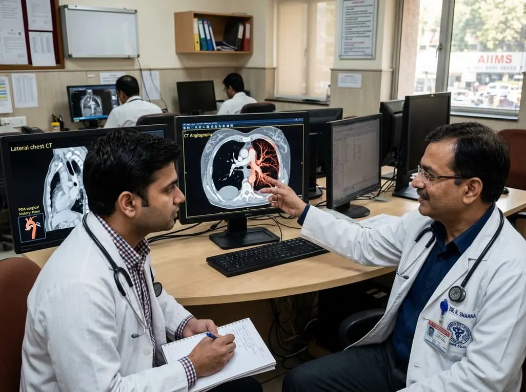
Congenital unilateral absence of the pulmonary artery can remain undetected until adulthood. This case report examines a young male presenting with hemoptysis after prior PDA surgery, highlighting the critical role of CT angiography in mapping systemic collaterals and guiding appropriate clinical intervention.
Today
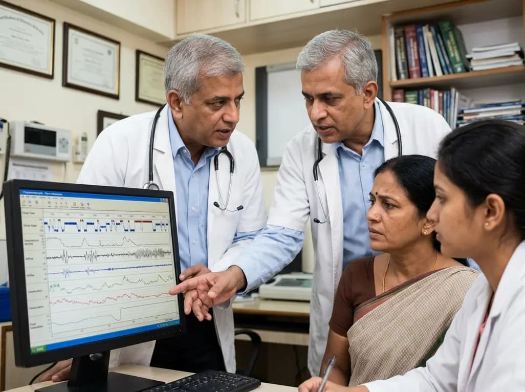
Perimenopausal obstructive sleep apnea often goes undetected due to atypical polysomnographic presentations and symptom overlap with menopause. This review details its neurocognitive mechanisms, polysomnography markers, and precision management strategies.
Today
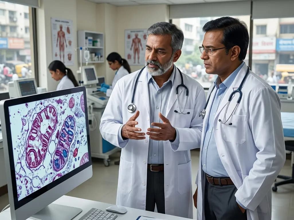
Mitochondrial deubiquitinases regulate organelle turnover by editing ubiquitin tags on damaged mitochondria. Discover how targeting these enzymes, notably USP30, offers therapeutic strategies for neurodegenerative, cardiovascular, and metabolic disorders.
Today

A rare case of a pediatric pathological femoral fracture caused by occult telangiectatic osteosarcoma demonstrates the risks of misdiagnosis after trauma, where intramedullary nailing prompted iatrogenic tumor dissemination and required modular total endoprosthetic reconstruction.
Today
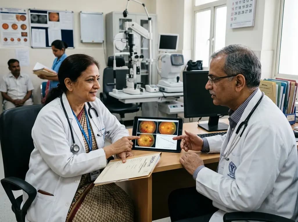
A comprehensive analysis of DRCRnet studies reveals that 15.1% of participants discontinue diabetic retinopathy clinical trials. Identifying baseline demographic and clinical predictors helps ophthalmologists and endocrinologists mitigate real-world treatment dropouts and protect long-term visual outcomes.
Today
