

Loading, please wait...
Loading, please wait...

Levodopa remains the cornerstone pharmacological therapy for motor symptoms in Parkinson's disease. However, clinicians frequently observe substantial variability in levodopa responsiveness across individual patients. While some individuals maintain excellent symptomatic relief over many years, others exhibit inadequate response or develop debilitating motor fluctuations. Traditionally, clinicians attributed these discrepancies primarily to advancing disease duration and progressive dopaminergic denervation. Yet, accumulating evidence reveals that heterogeneous biological mechanisms govern this pharmacological variability. To address these critical clinical questions, investigators evaluated multimodal neuroimaging signatures within the well-characterized PREDISTIM cohort of candidates undergoing deep brain stimulation. Understanding the precise neural substrates governing drug efficacy allows neurologists to move beyond empirical medication adjustments. Moreover, identifying objective imaging markers provides profound insights into patient selection for invasive therapies. Because clinical assessments alone can introduce significant subjectivity, quantitative neuroimaging techniques offer reliable windows into underlying circuit disruption. Consequently, distinguishing these diverse biological drivers enhances our ability to deliver tailored therapy. This investigative framework bridges the gap between symptomatic assessment and neurobiological mechanisms, thereby refining therapeutic expectations in complicated movement disorders.
Historically, medical practice categorized patients through a binary framework consisting of good responders and poor responders. However, recent data-driven methodologies challenge this oversimplified view. By employing unsupervised clustering algorithms that simultaneously incorporate dopaminergic responsiveness and disease duration, researchers uncovered three discrete phenotypic clusters. Interestingly, this classification mirrors findings previously observed in the Parkinson's Progression Markers Initiative cohort. Cluster 3 represents classic robust responders who exhibit significant motor improvement after drug administration. Conversely, cluster 1 comprises poor responders who experience negligible clinical benefit. Most notably, the analysis identified cluster 2 as an intermediate phenotype characterized by preserved pharmacological responsiveness despite lengthy disease duration. Therefore, clinical progression does not follow a strict linear pathway across all individuals. Instead, these findings establish that levodopa sensitivity operates across unique phenotypic trajectories. Neurologists should recognize that long disease duration does not automatically preclude meaningful pharmacological responsiveness. In addition, this tripartite categorization confirms that differential neurodegenerative processes dictate individual therapeutic outcomes. Recognizing these divergent patterns helps clinicians customize pharmacotherapy more effectively.
Multimodal magnetic resonance imaging delivers critical insights into structural alterations across these distinct clinical clusters. High-resolution T1-weighted structural scans demonstrated significant putaminal atrophy specifically within cluster 2 patients. Because the putamen receives extensive dopaminergic projections from the substantia nigra, structural volume loss within this region directly impacts sensorimotor processing. Nevertheless, these patients preserve meaningful therapeutic responsiveness, suggesting compensatory cortical mechanisms or localized preservation of postsynaptic dopamine receptors. In sharp contrast, patients in cluster 1 showed preserved putaminal volume but displayed marked grey matter loss in the temporo-parietal operculum and inferior frontal cortex. These specific cortical zones contribute extensively to motor planning, cognitive control, and multisensory integration. Thus, poor dopaminergic responsiveness reflects extensive extrastriatal structural damage rather than isolated basal ganglia failure. Consequently, motor deficits in non-responders stem from disrupted large-scale networks that dopamine replacement cannot overcome. These structural observations emphasize the need for comprehensive radiological assessments prior to surgical interventions. Furthermore, identifying regional volume deficits allows physicians to predict which motor signs will resist standard pharmacological escalations.
In addition to macroscopic volume loss, microenvironmental tissue changes significantly influence therapeutic outcomes. Utilizing multi-echo fast gradient-echo magnetic resonance sequences, researchers successfully quantified regional iron accumulation. Cluster 1 patients exhibited markedly elevated iron deposition within the substantia nigra pars compacta and the globus pallidus internus. Excessive iron accumulation promotes severe oxidative stress, mitochondrial failure, and secondary neuroinflammation. Moreover, this metallic burden correlates with progressive neuronal injury and irreversible microstructural destruction. When substantial iron accumulates within the globus pallidus internus, normal basal ganglia outflow suffers profound destabilization. As a consequence, exogenous levodopa cannot adequately restore physiological firing patterns across the thalamocortical pathway. Furthermore, elevated iron levels within these deep subcortical nuclei suggest aggressive localized degeneration. Therefore, susceptibility-weighted imaging and quantitative susceptibility mapping serve as valuable diagnostic adjuncts for evaluating refractory motor symptoms. By detecting microenvironmental degradation before gross tissue destruction becomes obvious, clinicians gain crucial diagnostic clarity. Ultimately, these advanced sequences differentiate primary striatal dysfunction from widespread basal ganglia pathology.
These radiological discoveries provide indispensable guidance for deep brain stimulation workflows. Preoperative evaluation traditionally relies upon the acute levodopa challenge test to confirm surgical candidacy. However, performing drug withdrawal and acute rechallenge often triggers severe patient discomfort and potential complications. Integrating objective magnetic resonance markers streamlines this screening process significantly. Specifically, patients with marked cortical atrophy and severe pallidal iron overload derive less benefit from conventional pharmacotherapy and may present altered surgical outcomes. Conversely, patients in cluster 2 maintain high responsiveness despite putaminal volume changes, making them favorable surgical candidates. In addition, recognizing extrastriatal degenerative patterns assists neurosurgeons and neurologists in counseling families regarding realistic postoperative expectations. Clinicians must realize that subthalamic stimulation primarily relieves symptoms that respond positively to levodopa. Consequently, identifying non-dopaminergic cortical degeneration prevents inappropriate surgical selection and avoids procedural dissatisfaction. Furthermore, these imaging biomarkers support the formulation of customized targeting strategies tailored to specific circuit vulnerabilities. Multimodal imaging thus enhances safety and clinical precision in neuromodulation.
Implementing multimodal neuroimaging signatures into routine clinical workflows promises to revolutionize personalized medicine for movement disorders. While specialized academic centres currently lead these technological applications, standardizing imaging protocols will expand access across general neurology practices. Clinicians can utilize routine brain magnetic resonance imaging not merely to exclude secondary parkinsonism, but also to evaluate regional atrophy and iron deposition quantitatively. Moreover, recognizing that levodopa response variability reflects distinct neurobiological subtypes enables proactive therapeutic planning. Rather than escalating levodopa dosages to intolerable levels in cluster 1 patients, clinicians can prioritize non-dopaminergic options, physical rehabilitation, and multidisciplinary care early. Similarly, identifying cluster 2 patients reassures physicians that intensive dopaminergic optimization remains effective even late in the disease course. Therefore, combining clinical assessments with structural and susceptibility-sensitive imaging markers creates an actionable management matrix. Moving forward, collaborative multicentre studies must validate these imaging thresholds across diverse global populations. In doing so, modern neurology takes another decisive step toward truly individualized, biology-driven care for Parkinson's disease.
Multimodal MRI combines high-resolution structural imaging with iron-sensitive sequences to detect regional cortical atrophy and subcortical iron accumulation. Consequently, it reveals whether persistent motor deficits stem from localized striatal degeneration or broader network destruction, explaining why patients with similar clinical disease stages respond differently to dopaminergic medication.
Deep brain stimulation outcomes depend heavily upon baseline levodopa responsiveness. Stratifying patients into data-driven clusters identifies individuals with preserved dopa sensitivity despite extended disease duration. Moreover, detecting severe pallidal iron deposition and extrastriatal cortical atrophy warns clinicians of atypical circuit degeneration that may limit surgical benefits.
Currently, standard clinical therapies cannot reverse pathological iron deposition within the basal ganglia. However, detecting increased iron deposition in the substantia nigra and globus pallidus internus provides indispensable prognostic information. It signals aggressive localized neurodegeneration, guiding clinicians to avoid excessive levodopa escalations and focus on comprehensive management.
Disclaimer: This content is for informational and educational purposes only and should not be considered medical advice or substituted for professional clinical judgment. Diagnostic criteria, dosing, and therapeutic strategies should be customized to each patient. Refer to the latest local and national guidelines for clinical practice.
References

Read summarized clinical updates, watch expert medical content, and earn CME certifications right from your smartphone.
Open your smartphone camera to scan and install.





A multimodal MRI study in deep brain stimulation candidates demonstrates that levodopa responsiveness in Parkinson's disease reflects distinct neurobiological phenotypes, characterized by specific cortical atrophy and basal ganglia iron deposition patterns rather than simple disease progression.
Today

A landmark study reveals that 20 minutes of moderate aerobic exercise shields episodic memory encoding as effectively as a 90-minute nap after 30 hours of sustained wakefulness, offering clinicians and shift workers a viable neuroprotective strategy against acute sleep loss.
Today

Biotechnology researchers in India are developing an automated liquid biopsy platform to detect cervical malignancy from blood samples. Supported by the MAHA MedTech Mission, this initiative applies nanopore sequencing and piRNA biomarker detection to eliminate invasive speculum exams and expand rural screening.
Today
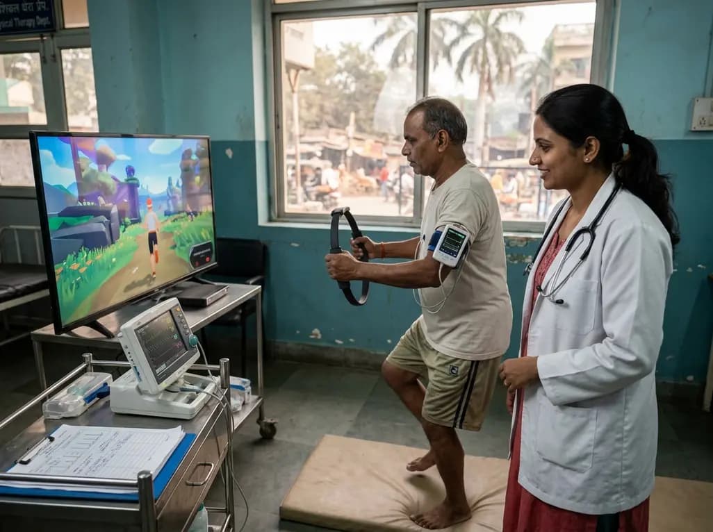
A clinical study evaluated Ring Fit Adventure in adults with chronic conditions, revealing that exergaming achieves moderate-to-vigorous exercise intensity while maintaining hemodynamic stability, high enjoyment, and zero adverse events.
Today

A retrospective cohort study reveals that selecting the touched vertebra as the lowest instrumented vertebra in thoracic adolescent idiopathic scoliosis yields a 20.8% distal add-on rate, driven by remnant motion segments and axial rotational discrepancies.
Today
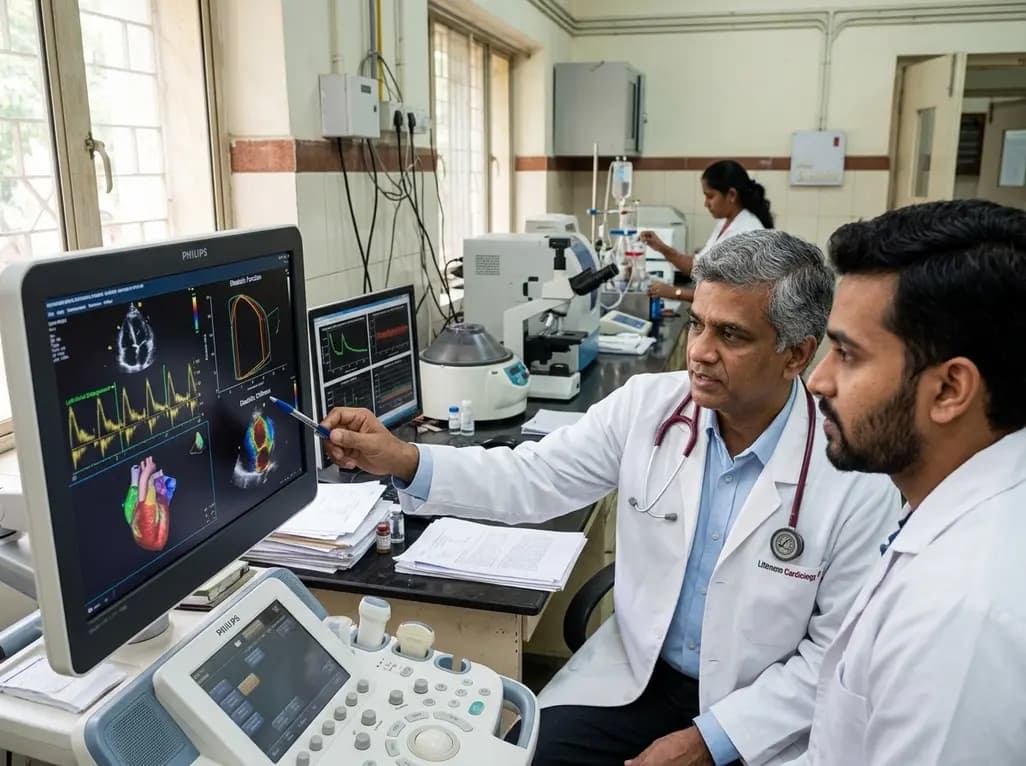
A study evaluates a cardiometabolic HFpEF model combining metabolic stress with chronic mineralocorticoid excess. The findings reveal profound diastolic stiffness, endothelial dysfunction, and pulmonary hypertension, illuminating key pathophysiological mechanisms for targeted cardiopulmonary therapies.
Today
