.webp&w=384&q=75)

Loading, please wait...
Loading, please wait...

Modern oncology has markedly improved survival outcomes for patients living with intracranial neoplasms. However, clinicians increasingly confront the long-term cognitive and neurological consequences of cranial irradiation. Specifically, understanding radiation therapy brain changes in non-tumoral cerebral parenchyma remains critical for optimizing post-treatment quality of life. Ionizing radiation causes complex cellular injury within healthy neural tissues, affecting both white matter architecture and cortical gray matter. Advanced quantitative magnetic resonance imaging now allows investigators to track these progressive alterations over time. Consequently, identifying early and late neuroarchitectural shifts enables clinicians to tailor radiotherapy plans more effectively.
Postoperative cranial irradiation delivers substantial therapeutic benefits against residual glioma cells. Nevertheless, ionizing beams inevitably traverse normal cerebral regions surrounding the resection cavity. Researchers sought to examine these healthy compartments longitudinally using advanced volumetric and diffusion MRI techniques. The study evaluated glioma cohorts undergoing conformal radiation doses ranging from 25 to 60 Gy. Clinicians gathered imaging data before treatment, in an early follow-up window within 8 months, and in a late phase spanning 9 to 16 months. Linear mixed-effects modeling allowed investigators to separate tumor effects from genuine radiation injury across cortical and subcortical structures.
As a result, investigators documented measurable reductions in regional brain volume and widespread cortical thinning over time. In addition, quantitative diffusion sequences showed clear alterations in apparent diffusion coefficient values across diverse gray matter sites. These metrics reflect microscopic perturbations before macrostructural necrosis appears on standard diagnostic scans. Furthermore, early detection enables timely rehabilitation before irreversible neurocognitive decline occurs. Therefore, quantitative neuroimaging provides an indispensable window into subclinical radiation neurotoxicity.
Diffusion-weighted imaging measures the Brownian motion of water molecules within cerebral microenvironments. When radiation damages parenchymal integrity, apparent diffusion coefficient values elevate significantly. In the recent study, post-treatment scans revealed notable diffusion increases in the ipsilateral cerebral hemisphere. Specifically, regions showing pronounced diffusion elevations included the cuneus, precuneus, parahippocampal cortex, and superior parietal lobules. The inferior parietal, paracentral, and pericalcarine cortices also displayed significant microstructural loosening.
Moreover, these elevations indicate substantial loss of cellular barriers and progressive enlargement of extracellular fluid spaces. Radiation induces localized endothelial apoptosis, disrupts the blood-brain barrier, and triggers chronic microglial activation. Consequently, neuroinflammation impairs synaptic arborization and diminishes oligodendrocyte survival. In addition, the ipsilateral hemisphere absorbs higher cumulative scatter doses due to proximity to the primary glioma bed. Thus, elevated diffusion metrics serve as sensitive early surrogates for subtle neurodegenerative remodeling. By recognizing these diffusion alterations, neuro-oncologists can anticipate regional functional vulnerabilities before profound clinical deficits emerge.
Radiation injury extends far beyond the immediate surgical perimeter. Indeed, statistical models controlling for age, sex, tumor grade, scanner variations, and timing revealed dose-dependent changes in distant areas. Most strikingly, apparent diffusion coefficient increases directly correlated with delivered radiation dose in contralateral structures. These sensitive regions included the contralateral middle temporal gyrus, pars opercularis, and lingual gyrus.
Therefore, cerebral parenchyma displays selective regional vulnerability even at moderate or low scatter doses. In particular, the middle temporal gyrus plays an essential role in semantic memory and language comprehension. Similarly, the pars opercularis contributes to expressive language fluency and motor planning networks. Meanwhile, the lingual gyrus processes higher-order visual cues and memory consolidation. Radiation damage to these contralateral processing centers can provoke insidious neurocognitive deficits that standard bedside testing often misses. Furthermore, these dose correlations demonstrate that scatter radiation exerts measurable biological consequences across both cerebral hemispheres. Clinicians must acknowledge that distant contralateral cortex does not remain an innocent bystander during focused cranial radiotherapy.
Ionizing radiation generates abundant reactive oxygen species that inflict severe oxidative stress on cerebral parenchyma. As a consequence, mature neurons and neural progenitor cells suffer chronic DNA damage. Radiation also targets endothelial cells within delicate cerebral capillaries, causing microvascular depletion and local ischemia. Consequently, microcirculatory breakdown increases blood-brain barrier permeability and incites persistent neuroinflammation. Activated astrocytes and microglia produce abundant pro-inflammatory cytokines, which perpetually suppress adult hippocampal neurogenesis.
Furthermore, radiation disrupts oligodendrocyte precursors, precipitating demyelination and axonopathy across vulnerable white matter tracts. Over time, progressive cellular attrition manifests as cortical thinning, ventricular enlargement, and measurable brain volume contraction. Similarly, extracellular matrix degradation expands interstitium, producing the marked diffusion elevations captured on quantitative neuroimaging. Although oncologists prioritize tumoricidal radiation delivery, this continuous biochemical cascade damages healthy gray and white matter. Thus, modern neuro-oncology must develop balanced approaches that combine tumor control with active parenchymal neuroprotection. Recognizing these pathophysiological pathways helps clinicians implement timely medical and radiological countermeasures.
These quantitative MRI insights demand meaningful changes in routine clinical radiation planning. Modern radiation oncology offers sophisticated technologies, including intensity-modulated radiotherapy and volumetric modulated arc therapy. Radiation oncologists can leverage these conformal methods to sculpt steep dose gradients around critical cortical regions. For example, clinicians already practice hippocampal-avoidance techniques to preserve verbal memory in brain metastasis treatment. Similarly, avoiding unnecessary dose exposure to the middle temporal and inferior frontal regions may safeguard essential language networks.
In addition, proton beam therapy provides distinct physical advantages through its sharp Bragg peak. Protons stop abruptly at designated target depths, sparing downstream cerebral parenchyma from unwanted exit doses. Moreover, pharmacological neuroprotectants and lifestyle interventions show immense promise in ongoing clinical trials. Agents such as memantine mitigate excitotoxicity, while tailored physical exercise stimulates neurotrophic factor release and cognitive reserve. Therefore, integrating quantitative MRI into routine follow-up protocols empowers neuro-oncology teams to track subclinical tissue degeneration. Through personalized dose constraints and proactive neuroprotective strategies, multidisciplinary teams can maintain glioma survival gains while preserving vital cognitive independence.
Radiation therapy induces oxidative stress, microvascular injury, and chronic neuroinflammation within healthy brain parenchyma. These biological cascades destroy oligodendrocyte precursors, disrupt endothelial barriers, and damage neuronal networks. Consequently, patients exhibit progressive cortical thinning, measurable tissue volume reduction, and increased water diffusivity across sensitive gray and white matter regions.
The apparent diffusion coefficient rises because radiation degrades cellular density and membrane integrity. Endothelial apoptosis, demyelination, and neuronal attrition expand the extracellular fluid compartment. As a result, water molecules move more freely through disrupted parenchymal matrices, producing elevated diffusion signals on quantitative MRI before gross structural necrosis becomes visible.
Clinicians can employ advanced conformal radiation technologies like intensity-modulated radiotherapy and proton therapy to sculpt sharp dose boundaries. Specifically sparing critical neurogenic structures, including the hippocampus and eloquent contralateral cortices, protects cognitive networks. In addition, neuroprotective medications like memantine and early cognitive rehabilitation substantially lessen post-treatment neurological deficits.
Disclaimer: This content is for informational and educational purposes only and should not be considered medical advice. Always consult a qualified healthcare provider for specific clinical concerns. Refer to the latest local and national guidelines for clinical practice.
References

Read summarized clinical updates, watch expert medical content, and earn CME certifications right from your smartphone.
Open your smartphone camera to scan and install.





A recent quantitative MRI study reveals that radiation therapy causes progressive structural brain changes, including cortical thinning and microstructural diffusion alterations in healthy tissue among glioma survivors. These findings emphasize the clinical need for dose-sparing neuroprotective techniques.
Today
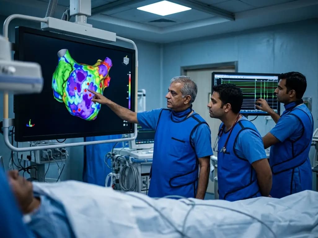
A study of 1,428 AF ablation patients reveals that over 60% of left atrial non-PV triggers arise within low-voltage areas, with anterior and roof triggers colocalizing entirely within fibrotic substrate. Identifying and ablating these sites is critical for long-term arrhythmia freedom.
Today

A comprehensive meta-analysis of over 278,000 women demonstrates that maternal atopic dermatitis significantly increases risks of premature rupture of membranes and low birth weight. Discover essential clinical insights on disease mechanisms, serial antenatal growth monitoring, and safe therapeutic interventions.
Today
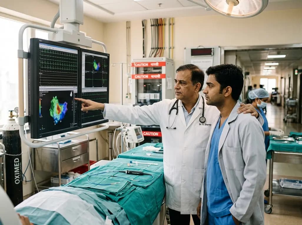
A pioneering study highlights the first United States experience using the FARAPOINT focal pulsed-field ablation catheter for refractory ventricular tachycardia. The catheter achieved complete acute non-inducibility across tested patients without procedural complications, marking a milestone in ventricular ablation.
Today

A biomechanical study shows that augmenting thin fascia lata grafts with a reinforced polyester patch doubles ultimate tensile strength and stiffness, matching the performance of thick grafts for superior capsule reconstruction while reducing donor-site morbidity.
Today

A cross-sectional study reveals that elevated METS-IR is independently associated with higher myopia odds in adolescents, showing biological synergy with obesity and highlighting the need for metabolic screening in pediatric eye care.
Today
