.webp&w=384&q=75)

Loading, please wait...
Loading, please wait...

Recurrent patellofemoral instability presents a complex diagnostic and reconstructive challenge for modern orthopaedic surgeons. Young athletes frequently sustain lateral patellar dislocations during cutting maneuvers. Consequently, clinicians must accurately identify underlying structural risk factors to prevent recurrent episodes. Historically, clinicians have relied on measurements such as trochlear dysplasia, patella alta, and coronal malalignment. However, traditional coronal indices often fail to capture functional patellofemoral engagement during early knee flexion. Because early flexion represents the most vulnerable arc of motion, precise assessment remains vital. In patellofemoral instability, the extensor mechanism often displaces laterally relative to the femoral trochlea. Therefore, identifying accurate anatomic parameters guides appropriate surgical interventions. Although medial patellofemoral ligament reconstruction restores medial restraint, persistent extensor malalignment causes graft failure. As a result, surgical planning demands reliable imaging parameters that separate true pathology from benign anatomic variation. Recent investigation focuses on how well the bony trochlea physically contains the extensor mechanism. Specifically, researchers introduced the patellar tendon-lateral trochlear ridge distance to evaluate lateral extensor drift. This innovative parameter addresses key limitations present in established imaging protocols. Thus, sports medicine specialists are re-evaluating traditional diagnostic paradigms to optimize patient care.
For decades, surgeons have used the tibial tubercle-trochlear groove distance to measure lateralization. Furthermore, clinicians often use the tibial tubercle-posterior cruciate ligament metric as an alternative reference. Both measurements quantify the lateral displacement of the tibial tuberosity on cross-sectional imaging. However, neither index directly assesses the soft tissue extensor mechanism. Instead, they focus solely on bony landmarks that vary across patient cohorts. Moreover, rotational alignment changes between the femur and tibia alter these measurements substantially. Knee flexion angle during magnetic resonance imaging also shifts values unpredictably. In addition, the tibial tubercle-trochlear groove distance demonstrates high sensitivity but suffers from limited specificity. Consequently, many asymptomatic individuals exhibit borderline or elevated values. This diagnostic overlap creates uncertainty when deciding whether to perform a tibial tubercle osteotomy. Unnecessary bony realignment introduces significant surgical risks, including nonunion and fracture. Therefore, orthopaedic specialists require discriminative imaging markers before committing patients to osteotomy. While the posterior cruciate ligament serves as a stable tibial landmark, it cannot evaluate patellar engagement. Thus, relying strictly on traditional parameters may misguide operative strategies. Clinicians clearly need imaging metrics that correlate directly with true patellofemoral tracking abnormalities.
To overcome the limitations of bony tuberosity measurements, investigators developed the patellar tendon-lateral trochlear ridge distance. This parameter measures the lateral edge of the patellar tendon relative to the lateral trochlear ridge. Because the patellar tendon represents the actual functional vector of the extensor mechanism, it reflects true tracking. Specifically, the lateral trochlear ridge acts as the critical bony wall preventing lateral subluxation. When the patellar tendon drifts lateral to this bony barrier, patellar engagement fails during early flexion. Consequently, the extensor mechanism lacks bony containment when quadriceps forces increase. In contrast to conventional indices, this metric integrates soft tissue orientation with functional trochlear geometry. Furthermore, magnetic resonance imaging easily identifies both the patellar tendon and the lateral trochlear ridge. Axial slices at the proximal trochlea establish bony references, while tendon margins provide soft tissue borders. Additionally, this approach eliminates inaccuracies caused by varying knee rotation during scanning. Therefore, the measurement provides a reproducible assessment of patellar containment within the intercondylar groove. By evaluating functional anatomy rather than isolated bony points, clinicians gain deeper insight into patellofemoral mechanics. Consequently, this measurement serves as an intuitive indicator of dislocation risk.
Recent clinical studies directly compare the diagnostic performance of these competing imaging measurements. In a propensity-matched case-control study, researchers evaluated adolescent patients undergoing primary patellofemoral stabilization. Notably, both the traditional tuberosity and novel tendon measurements demonstrated excellent reliability. However, substantial differences emerged regarding diagnostic specificity. The patellar tendon-lateral trochlear ridge distance demonstrated markedly higher specificity for identifying recurrent patellofemoral instability. In this comparative cohort, patients with instability exhibited an average distance difference of seven millimeters compared to healthy controls. Furthermore, receiver operating characteristic analyses confirmed superior area under the curve values. While traditional tuberosity indices captured most unstable knees, they frequently produced false positives in matched controls. Conversely, the tendon-to-ridge distance accurately distinguished pathological extensor lateralization without misclassifying normal knees. Consequently, this elevated specificity reduces the risk of unnecessary surgical overcorrection. Moreover, the statistical robustness of this metric held true across both male and female cohorts. These findings indicate that soft tissue containment metrics offer superior diagnostic precision. Therefore, radiologists and surgeons can utilize this threshold with greater diagnostic confidence.
Integrating the patellar tendon-lateral trochlear ridge metric into clinical workflows substantially improves surgical planning. In routine practice, orthopaedic teams must decide between isolated soft tissue reconstruction and combined bony realignment. Isolated reconstruction of the medial patellofemoral ligament suffices for patients with normal coronal tracking. However, performing isolated ligament reconstruction in the presence of severe extensor malalignment invites recurrent instability. Conversely, performing a tibial tubercle osteotomy in a patient with normal extensor tracking creates unnecessary surgical morbidity. Therefore, using a highly specific measurement prevents both undertreatment and overtreatment. When magnetic resonance imaging demonstrates an elevated tendon-to-ridge distance, surgeons can confidently recommend bony realignment. Additionally, this measurement aids in postoperative assessment to verify that surgical transfer achieved appropriate extensor recentering. Furthermore, sports medicine teams can incorporate this metric into standardized knee imaging templates. As protocols advance, automated artificial intelligence tools may measure this distance routinely. Ultimately, adopting this parameter refines patient selection, optimizes surgical outcomes, and protects long-term joint health. Consequently, clinicians should consider incorporating this reliable measurement into their standard preoperative evaluations.
The patellar tendon-lateral trochlear ridge distance evaluates the functional extensor mechanism directly rather than relying solely on bony landmarks. Traditional measurements assess the position of the tibial tuberosity relative to the femoral groove. However, they do not account for soft tissue extensor drift or dynamic knee rotation. In contrast, this tendon-based metric measures actual patellar tendon lateralization beyond the protective lateral trochlear ridge, providing superior diagnostic specificity for true patellofemoral instability.
Surgeons should order magnetic resonance imaging following any acute patellar dislocation or in patients presenting with recurrent instability symptoms. Cross-sectional imaging accurately identifies chondral shear injuries, medial patellofemoral ligament tears, and underlying trochlear dysplasia. Furthermore, magnetic resonance scans allow precise quantification of coronal malalignment parameters without radiation exposure. Clinicians can evaluate the patellar tendon-lateral trochlear ridge distance alongside traditional bony measurements to determine whether surgical stabilization requires concurrent bony realignment.
An elevated tendon-to-ridge distance indicates significant lateral displacement of the extensor vector, impairing trochlear engagement during early flexion. When this distance exceeds established diagnostic thresholds, isolated soft tissue reconstruction frequently fails due to uncorrected biomechanical loads. Therefore, surgeons utilize this measurement to identify patients who genuinely require tibial tubercle osteotomy. Combining medial patellofemoral ligament reconstruction with medializing tubercle transfer restores physiological tracking, relieves lateral patellar stress, and prevents persistent instability.
Disclaimer: This content is for informational and educational purposes only. It is not intended to be a substitute for professional medical advice, diagnosis, or treatment. Always seek the advice of your physician or other qualified health provider with any questions you may have regarding a medical condition. Refer to the latest local and national guidelines for clinical practice.
References

Read summarized clinical updates, watch expert medical content, and earn CME certifications right from your smartphone.
Open your smartphone camera to scan and install.





A recent comparative study shows patellar tendon-lateral trochlear ridge (PT-LTR) distance offers greater specificity than TT-TG for patellofemoral instability. This soft-tissue metric improves diagnostic accuracy and guides surgical decision-making for medial patellofemoral ligament and tubercle procedures.
Today
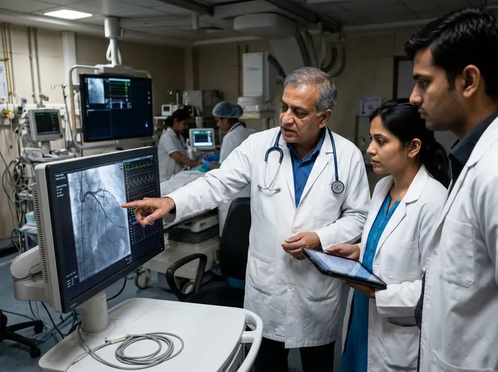
New evidence demonstrates that the atherogenic index of plasma (AIP) is an independent predictor of the slow flow/no-reflow phenomenon in acute myocardial infarction patients undergoing emergency PCI, offering clinicians a valuable tool for early risk stratification and targeted microvascular protection.
Today
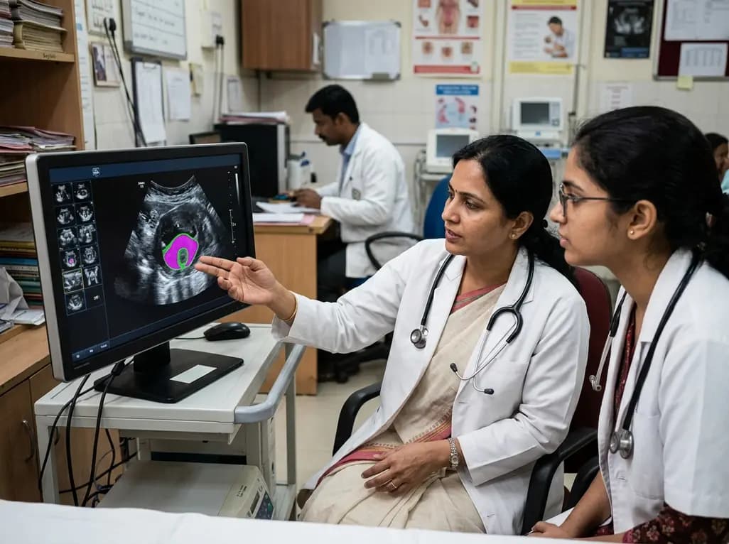
New evidence evaluates MoUNets, a static-weighted ensemble of UNet experts for automated levator hiatus segmentation in pelvic floor ultrasound. Learn how deep learning architectures perform in pelvic organ prolapse diagnostics and clinical practice.
Today
Managing destructive Citrobacter koseri endocarditis with aortocavitary fistula requires meticulous antiplatelet bridging, extensive debridement, and complex redo aortic root and RVOT reconstruction.
Today

A cross-sectional study reveals that elevated METS-IR is independently associated with higher myopia odds in adolescents, showing biological synergy with obesity and highlighting the need for metabolic screening in pediatric eye care.
Today
