

Loading, please wait...
Loading, please wait...

The human parietal lobe functions as a complex, highly integrated neurocognitive crossroads. It orchestrates language comprehension, somatosensory integration, visuospatial processing, and executive operations. When primary intrinsic neoplasms arise in this eloquent region, clinicians face substantial therapeutic challenges. Specifically, adult patients with parietal glial tumors often manifest subtle cognitive impairments before surgical intervention. Understanding these baseline deficits is essential for maximizing cytoreduction while protecting long-term quality of life. Recent clinical evidence offers valuable descriptive insights into how these neoplasms compromise distinct neurological networks.
Primary brain tumors situated within the parietal cortex rarely present solely with overt sensorimotor paralysis. Instead, individuals with parietal glial tumors frequently harbor profound yet covert neuropsychological deficits before surgical intervention. In a landmark multicenter investigation spanning two decades, researchers evaluated adult patients with newly diagnosed parietal lesions within fifteen days of diagnosis. Histologically, WHO grade 2 oligodendrogliomas represented the predominant pathology, followed by other diffuse glial and glioneuronal subtypes. Notably, comprehensive neuropsychological testing revealed that executive dysfunction dominated the clinical picture across the cohort. Because bedside mini-mental screens lack diagnostic granularity, standard clinical exams routinely fail to detect these impairments. Consequently, patients often experience significant difficulties in professional and social environments long before undergoing surgical resection. Diffuse infiltration along subcortical associative tracts disrupts distributed functional networks well beyond visible magnetic resonance imaging lesion boundaries. Therefore, neurosurgeons and neuro-oncologists must recognize that cortical infiltration silently undermines complex higher-order cognitive processing. Establishing a structured baseline assessment battery enables clinicians to document existing deficits accurately. Ultimately, this granular baseline provides a critical benchmark for postoperative comparison and longitudinal survivorship care.
Laterality plays a pivotal role in dictating the phenotypic cognitive presentation of parietal lesions. In descriptive analyses, right-sided tumors demonstrated prominent impairments in abstract reasoning, affecting sixty percent of tested individuals. Additionally, over forty percent of patients with right parietal tumors exhibited deficits in learning and memory recall. Working memory and cognitive flexibility also showed deficits in nearly forty percent of these cases. In contrast, left-sided parietal lesions generated a divergent pattern dominated by executive and linguistic vulnerabilities. For instance, over sixty-three percent of patients with left parietal neoplasms demonstrated impaired cognitive flexibility. Furthermore, working memory deficits occurred in more than half of left-sided cases, while forty percent suffered from reduced mental inhibition. Although statistical comparisons between hemispheres did not attain formal significance due to sample size constraints, the descriptive divergence remains clinically instructive. These findings illustrate that both hemispheres contribute substantially to distinct facets of executive control. Therefore, neuro-oncologists must not assume that cognitive compromise is confined to the dominant hemisphere. Instead, clinicians should anticipate specific cognitive vulnerabilities depending on hemispheric laterality, designing tailored neuropsychological batteries that assess both dominant and non-dominant cortical processing.
Beyond hemispheric laterality, precise anatomical sublocalization dictates the severity and breadth of cognitive impairment. Anatomists divide the parietal cortex into several functional subregions, including the postcentral gyrus, superior parietal lobule, supramarginal gyrus, angular gyrus, and precuneus. Among these diverse structures, the left precuneus demonstrated the most widespread cognitive disruption in recent observational data. Specifically, tumors involving the left precuneus caused impairments that exceeded predefined descriptive thresholds across numerous neurocognitive domains. In contrast, other parietal subregions exhibited more selective or inconsistent neuropsychological deficits. Neuroscience explains this vulnerability through structural and functional connectivity. The precuneus serves as a core metabolic and architectural hub within the default mode network and the frontoparietal control network. Consequently, even low-grade infiltrating neoplasms within the precuneus can disrupt global network communication. Lesions in this area impair episodic memory retrieval, visuospatial representation, and conscious information integration. Therefore, neurosurgeons planning resections near the medial parietal cortex must account for the extensive network vulnerability of the precuneus. Recognizing these subregional distinctions allows clinical teams to anticipate perioperative functional risks more effectively.
Modern neuro-oncology demands an optimal balance between maximizing the extent of tumor resection and preserving functional independence. Historically, surgical strategies prioritized the avoidance of primary motor or sensory deficits. However, modern clinical practice recognizes that higher-order cognitive deficits equally diminish patient autonomy, emotional well-being, and return to employment. Because parietal tumors frequently compromise cognitive flexibility, memory consolidation, and executive functioning, surgical resection strategies require advanced functional planning. Awake craniotomy with intraoperative electrical mapping represents the gold standard for navigating eloquent parietal subregions. In addition to conventional motor and somatosensory testing, surgical protocols should incorporate tailored intraoperative cognitive testing. For example, neurosurgeons can assess working memory, spatial orientation, calculation, and mental inhibition in real time during subcortical tumor dissection. Furthermore, integrating tractography and functional magnetic resonance imaging helps map critical subcortical white matter bundles, such as the superior longitudinal fasciculus and arcuate fasciculus. By combining preoperative neurocognitive profiles with dynamic intraoperative testing, neurosurgical teams minimize permanent postoperative cognitive decline. Consequently, patients achieve maximal oncological cytoreduction while maintaining their higher cognitive faculties.
Managing parietal glial and glioneuronal tumors requires comprehensive coordination across a multidisciplinary care team. Neurosurgeons, neuro-oncologists, clinical neuropsychologists, and rehabilitation specialists must collaborate from initial radiological diagnosis through long-term survivorship. Early preoperative neuropsychological evaluation provides an indispensable foundation for this multidisciplinary workflow. First, early testing alerts clinicians to subtle executive dysfunction that might compromise treatment adherence or informed consent comprehension. Second, baseline cognitive assessments inform tailored neuro-rehabilitation plans before surgical trauma occurs. Clinicians can introduce cognitive compensatory strategies immediately following surgical resection. Third, longitudinal cognitive tracking allows oncologists to distinguish between disease progression, surgical morbidity, and radiation-induced neurotoxicity. When clinicians initiate systemic therapies or radiotherapy, preexisting cognitive vulnerabilities require vigilant monitoring. Moreover, incorporating quantitative lesion mapping and advanced behavioral analytics into clinical trials will refine future therapeutic protocols. Ultimately, adopting a standardized, network-centered cognitive evaluation framework transforms brain tumor management. This holistic approach ensures that modern neuro-oncological treatment safeguards both overall survival and functional quality of life.
Baseline evaluation identifies subtle functional deficits that standard bedside exams frequently overlook. Moreover, comprehensive testing establishes an objective neurological reference point before surgical intervention. Consequently, surgical teams can tailor intraoperative functional mapping, optimize safe tumor resection boundaries, and proactively design personalized postoperative neuro-rehabilitation pathways to preserve patient independence.
While both hemispheres share executive dysfunction, their descriptive patterns diverge meaningfully. Right parietal lesions frequently disrupt abstract reasoning and spatial recall. In contrast, left parietal lesions commonly impair cognitive flexibility, working memory, and mental inhibition. Therefore, multidisciplinary teams should customize pre-surgical testing batteries based on tumor laterality to detect distinct cognitive vulnerabilities.
The precuneus serves as a central structural hub within default mode and frontoparietal networks. As a result, oncological lesions or surgical manipulation in this subregion disrupt multiple cognitive domains simultaneously. Patients often experience complex deficits across memory, mental processing, and executive control, necessitating meticulous intraoperative cortical and subcortical functional mapping.
Disclaimer: This content is for informational and educational purposes only and does not constitute medical advice, diagnosis, or treatment regimens. Healthcare professionals must exercise independent clinical judgment when evaluating individual patient cases and treatment strategies. Refer to the latest local and national guidelines for clinical practice.
References
Mallereau CH et al. Preoperative neuropsychological profiles in adult parietal glial and glioneuronal tumors: A retrospective descriptive study. Neurosurg Rev. 2026 Oct 19. doi: 10.1007/s10143-026-04542-0. PMID: 42894068.
Boone M, et al. Prevalence and profile of cognitive impairment in adult glioma: a sensitivity analysis. J Neurooncol. 2016;129(2):275-282.
van Kessel E, et al. Presurgical predictors of early cognitive outcome after brain tumor resection in glioma patients. Neurooncol Adv. 2021;3(1):vdab109.

Read summarized clinical updates, watch expert medical content, and earn CME certifications right from your smartphone.
Open your smartphone camera to scan and install.





Adults with parietal glial and glioneuronal tumors exhibit significant preoperative executive dysfunction. A 20-year study reveals distinct neuropsychological profiles across laterality and subregions, particularly highlighting precuneus vulnerability and informing tailored surgical mapping.
Today

A large UK cohort study of over 3.9 million patients found no substantive difference in the risk of peripheral neuropathy between systemic fluoroquinolones and cephalosporins, providing clinical reassurance while affirming known risks such as tendon rupture.
Today

A novel high-permeation vesicle-in-gel platform enhances transdermal fulvestrant delivery, achieving sustained release, robust intradermal retention, and superior antitumor efficacy in breast cancer models while circumventing the drawbacks of painful intramuscular depot injections.
Today
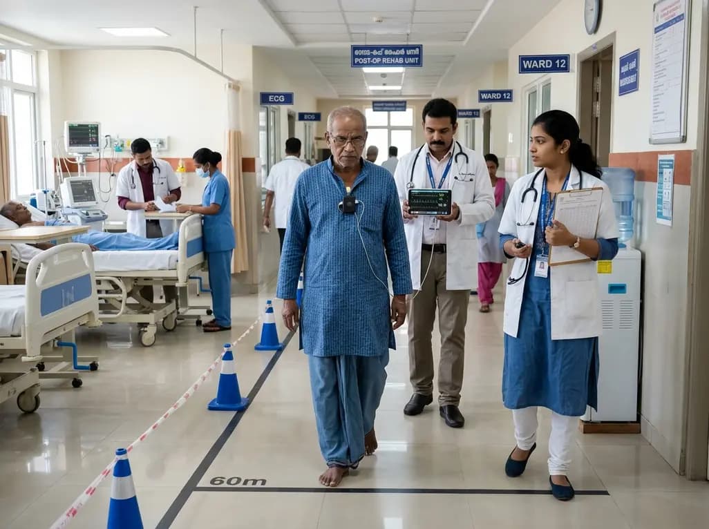
A recent study highlights the utility of measuring cardiac effort after surgery. In older, frail individuals undergoing cardiac surgery, combining continuous ECG with the six-minute walk test provides vital physiologic insights, demonstrating that heart rate responses mirror cycle ergometry.
Today
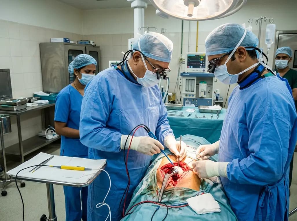
A systematic review and meta-analysis of 8 RCTs evaluated bipolar versus monopolar electrocautery in primary total hip and knee arthroplasty. Pooled data revealed no significant differences in haemoglobin drop, blood loss, or length of stay, concluding that routine bipolar electrocautery is not supported.
Yesterday

A critical review evaluates whether starch-polyphenol complexes meet structural and metabolic criteria to qualify as type 5 resistant starch. The proposed tiered evidence framework distinguishes true inclusion complexes from simple mixtures, clarifying mechanisms for glycemic modulation and gut fermentation.
Today
