.webp&w=384&q=75)

Loading, please wait...
Loading, please wait...

Genetic discoveries have transformed our understanding of frontotemporal dementia and amyotrophic lateral sclerosis over the past decade. Among the most frequent genetic etiologies are hexanucleotide mutations within the non-coding region of chromosome 9 open reading frame 72. Clinicians increasingly recognize that premanifest C9orf72 repeat expansions produce subtle neurofunctional alterations long before overt neurodegeneration occurs. Historically, many clinicians viewed early cognitive signs as generalized executive decline. However, recent evidence indicates that specific cognitive domains experience selective disruption during early stages. Understanding these early neurobiological vulnerabilities helps clinicians identify reliable prognostic markers and stage subclinical neurodegeneration effectively.
Hexanucleotide repeat mutations represent the primary monogenic cause linking frontotemporal lobar degeneration with amyotrophic lateral sclerosis. Carriers of premanifest C9orf72 repeat expansions often remain asymptomatic on standard bedside cognitive tests for decades. However, sophisticated neuropsychological assessments frequently reveal subtle episodic memory abnormalities. Researchers previously debated whether these memory deficits reflect diffuse cortical pathology or focal network breakdown. In addition, questions persisted regarding whether memory deficits affect all stimulus categories equally. Socioemotional stimuli carry profound biological and interpersonal significance. Consequently, researchers designed task-based functional neuroimaging protocols to isolate distinct memory stages. A critical investigation evaluated twenty-one premanifest mutation carriers alongside twenty-four demographically matched healthy control participants. Investigators instructed all participants to encode complex images comprising emotional faces, neutral faces, and inanimate houses. Subsequently, participants completed an immediate recognition retrieval test while undergoing high-resolution functional magnetic resonance imaging. This elegant paradigm allowed investigators to disentangle domain-general memory performance from domain-specific socioemotional memory processing. Furthermore, the protocol enabled detailed evaluation of both mass-univariate brain activations and multivoxel spatial patterns across cortical regions. Thus, the experimental setup provided unprecedented clarity regarding early subclinical neural changes.
The behavioral results revealed a striking and unexpected dissociation between stimulus categories. Specifically, premanifest carriers demonstrated significantly impaired recognition memory for human faces compared to healthy control participants. The statistical analysis showed marked impairment with a Mann-Whitney U value of 104 and high significance. However, premanifest carriers exhibited completely preserved recognition memory for inanimate houses. Their house recognition accuracy matched that of unaffected control individuals perfectly. Therefore, memory impairment during this premanifest phase is not a global episodic failure. Instead, the cognitive defect selectively targets socioemotional and interpersonal information. In clinical practice, subtle deficits in recognizing familiar faces or interpreting social cues often precede behavioral disturbances. Consequently, these findings validate patient families who report early, subtle social disconnects long before overt frontotemporal dementia develops. Furthermore, this dissociation suggests that specific neural networks mediating social cognition experience vulnerability before general hippocampal circuits deteriorate. Traditional screening tools such as the Mini-Mental State Examination often miss these fine domain-specific deficits. As a result, assessing social memory offers superior sensitivity for detecting the earliest cognitive manifestations of mutation carrier status. Clinicians must recognize that normal performance on standard object memory tests does not rule out socioemotional decline.
Functional neuroimaging during the initial memory encoding phase revealed distinct neural signatures in premanifest carriers. While viewing facial stimuli, premanifest individuals showed altered multivoxel activation patterns within the bilateral anterior insula. Advanced pattern classification algorithms successfully separated mutation carriers from healthy controls with classification accuracy exceeding seventy-six percent. Importantly, this statistical classification achieved robust significance across whole-brain and region-of-interest analyses. The anterior insula functions as an indispensable processing hub within the human brain. Specifically, it integrates autonomic physiological sensations with socioemotional appraisal and salient environmental stimuli. When healthy individuals encounter expressive faces, the anterior insula coordinates emotional decoding and directs cognitive resources toward encoding. However, in premanifest carriers, aberrant voxel-level activity patterns disrupt this crucial socioemotional evaluation. Consequently, the brain fails to establish strong, distinctive memory traces for human faces. In contrast, neural representations during house encoding remained largely preserved across groups. Furthermore, univariate analysis confirmed that this disruption did not reflect total neuronal loss or complete signal absence. Instead, it represented disorganized spatial organization across microcircuits within the anterior insula. Thus, functional insular reorganization serves as an early indicator of mutant gene activity before gross structural atrophy appears on anatomical scans.
The research team also identified profound neurofunctional disruptions during the memory retrieval phase. When attempting to recognize previously presented faces, premanifest carriers exhibited exaggerated neural responses in the right anterior insula. Specifically, univariate analyses revealed hyperactivation when comparing encoded target faces against novel foil faces. This heightened signal reached statistical significance with a Mann-Whitney U value of 394. Researchers interpret this excessive activation as compensatory recruitment attempting to overcome poorly organized initial encoding traces. Moreover, the investigators evaluated encoding-retrieval neural similarity across distributed functional brain networks. Successful episodic recollection normally relies on reactivating the exact neural patterns present during initial encoding. However, premanifest mutation carriers displayed significantly reduced neural pattern similarity between encoding and retrieval within the salience network. This reduction proved highly specific to facial stimuli and reached significance with a U value of 120. In contrast, pattern reinstatement for inanimate house stimuli showed no network-level deterioration. Therefore, the salience network suffers selective functional decoupling when handling socioemotional memories. Additionally, the amygdala and interconnected cingulate circuits exhibited parallel reductions in pattern reinstatement fidelity. Consequently, the brain struggles to verify whether an encountered face is familiar or novel, producing measurable recognition deficits.
These neuroimaging and behavioral findings offer substantial insights for clinical neurology and geriatric psychiatry. Historically, diagnosing frontotemporal spectrum disorders relied heavily on macrostructural magnetic resonance imaging showing visible lobar atrophy. Nevertheless, macrostructural atrophy represents late-stage irreversible neurodegeneration that offers poor opportunities for therapeutic rescue. Identifying functional alterations in the salience network provides clinicians with a much earlier physiological window. Because the anterior insula deteriorates earliest in behavioral variant frontotemporal dementia, tracking its functional integrity allows precise preclinical staging. Furthermore, multidimensional pattern analysis surpasses conventional univariate fMRI in detecting discreet neuronal stress. Multivoxel pattern abnormalities emerge years before volumetric loss appears on automated cortical thickness measurements. Thus, clinicians can potentially utilize task-based fMRI paradigms as objective diagnostic supplements when evaluating asymptomatic gene carriers. In clinical trial settings, functional connectivity and multivoxel metrics can serve as sensitive pharmacodynamic biomarkers. For instance, therapies aiming to reduce toxic dipeptide repeats could demonstrate efficacy by normalizing insular response patterns. Moreover, understanding that memory deficits in these carriers stem from salience network disruption helps differentiate them from Alzheimer disease. Unlike typical amnestic Alzheimer pathology centered on hippocampal storage failure, this disorder features impaired salience-driven encoding and retrieval verification.
Translating these specialized imaging findings into regular outpatient practice requires thoughtful cognitive profiling. Physicians caring for families affected by hereditary neurodegenerative conditions should screen for subtle changes in social cognition. In addition to testing standard verbal recall, clinicians should evaluate face recognition and emotional expression perception. Asking structured questions about subtle social withdrawal or misidentifying familiar acquaintances can uncover early premanifest symptoms. Additionally, genetic counseling plays an essential role when discussing these complex issues with at-risk relatives. Confirming carrier status demands comprehensive pre-test and post-test genetic counseling conducted by specialized multidisciplinary teams. Because effective disease-modifying therapies are currently undergoing global clinical trials, establishing accurate baseline profiles is critical. Neuroimaging centers should consider incorporating advanced resting-state or task-based fMRI protocols into dedicated research registries. Furthermore, prospective longitudinal monitoring will help determine the exact temporal latency between insular pattern disruption and overt clinical phenoconversion. Clinicians should maintain close collaboration between cognitive neurologists, neuropsychologists, and neuroimaging experts to support vulnerable families. Ultimately, identifying early biomarkers in premanifest carriers will enable timely therapeutic interventions before irreversible neuronal damage occurs.
Premanifest carriers experience selective memory challenges primarily involving socioemotional stimuli, such as remembering faces and social interactions. In contrast, domain-general memory for inanimate objects and spatial locations remains intact. These subtle difficulties stem from early functional disruptions within the brain's salience network, long before prominent dementia symptoms appear.
The anterior insula serves as a central hub within the salience network, integrating emotional awareness with sensory processing. In behavioral variant frontotemporal dementia and related spectrum disorders, insular microcircuits exhibit early neurochemical and metabolic susceptibility to toxic repeat expansions, leading to altered spatial activation patterns during socioemotional encoding.
Standard screening tests like the Mini-Mental State Examination generally fail to detect these subtle deficits because they evaluate broad orientation, language, and simple recall. Detecting premanifest social memory decline requires specialized neuropsychological tests focused on facial recognition, emotional appraisal, or sensitive task-based functional neuroimaging paradigms assessing insular pattern similarity.
Disclaimer: This content is for informational and educational purposes only... Refer to the latest local and national guidelines for clinical practice.
References

Read summarized clinical updates, watch expert medical content, and earn CME certifications right from your smartphone.
Open your smartphone camera to scan and install.





A groundbreaking fMRI study reveals that individuals with premanifest C9orf72 repeat expansions develop selective socioemotional memory deficits linked to salience network dysfunction, particularly in the anterior insula, well before the clinical onset of frontotemporal dementia or amyotrophic lateral sclerosis.
Today
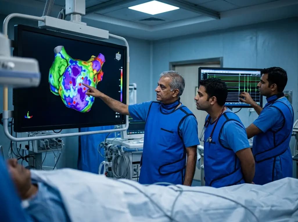
A study of 1,428 AF ablation patients reveals that over 60% of left atrial non-PV triggers arise within low-voltage areas, with anterior and roof triggers colocalizing entirely within fibrotic substrate. Identifying and ablating these sites is critical for long-term arrhythmia freedom.
Today

A comprehensive meta-analysis of over 278,000 women demonstrates that maternal atopic dermatitis significantly increases risks of premature rupture of membranes and low birth weight. Discover essential clinical insights on disease mechanisms, serial antenatal growth monitoring, and safe therapeutic interventions.
Today

A pioneering study highlights the first United States experience using the FARAPOINT focal pulsed-field ablation catheter for refractory ventricular tachycardia. The catheter achieved complete acute non-inducibility across tested patients without procedural complications, marking a milestone in ventricular ablation.
Today

A biomechanical study shows that augmenting thin fascia lata grafts with a reinforced polyester patch doubles ultimate tensile strength and stiffness, matching the performance of thick grafts for superior capsule reconstruction while reducing donor-site morbidity.
Today

A cross-sectional study reveals that elevated METS-IR is independently associated with higher myopia odds in adolescents, showing biological synergy with obesity and highlighting the need for metabolic screening in pediatric eye care.
Today
