.webp&w=384&q=75)

Loading, please wait...
Loading, please wait...
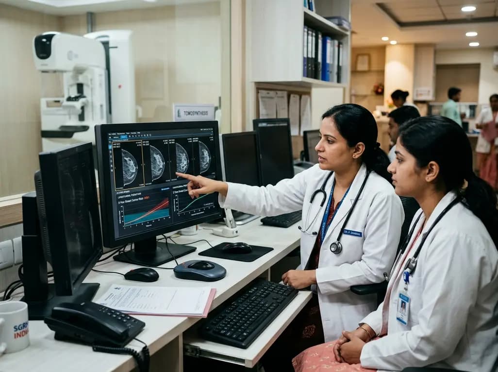
Accurate long-term breast cancer risk prediction remains a cornerstone of preventative oncology and personalized healthcare. Traditionally, risk estimation tools have relied heavily on clinical questionnaires, demographic factors, and single-timepoint imaging modalities such as full-field digital mammography. However, as medical technology evolves, digital breast tomosynthesis has progressively replaced standard two-dimensional mammography in modern screening programs. Despite its widespread adoption, the full predictive capability of sequential, three-dimensional imaging across multiple visits has remained underutilized until recently.
By leveraging deep learning algorithms, researchers are now tapping into the wealth of volumetric data embedded within longitudinal sequential scans. Evaluating subtle volumetric changes over time offers unprecedented insights into tissue dynamics, density alterations, and early parenchymal subtle shifts. Consequently, AI-driven breast cancer risk prediction models using longitudinal digital breast tomosynthesis provide a far more dynamic and comprehensive profile of a patient's long-term oncologic risk than static single-instance screening.
For decades, conventional full-field digital mammography served as the global standard for routine breast cancer screening. While effective, standard two-dimensional mammography suffers from tissue overlapping, which frequently masks underlying lesions or falsely simulates structural abnormalities. Digital breast tomosynthesis mitigates this limitation by reconstructing multiple thin quasi-three-dimensional slices. Consequently, tomosynthesis significantly improves diagnostic accuracy, enhances cancer detection rates, and lowers recall rates in diverse patient cohorts.
Nevertheless, early risk algorithms like the Tyrer-Cuzick model or single-scan AI architectures only analyze snapshot data from a single screening visit. This single-point evaluation misses longitudinal tissue trends, such as subtle asymmetric density progression over sequential years. Integrating serial examinations allows deep learning algorithms to track individual baseline variations over time. Therefore, longitudinal modeling captures cumulative architectural alterations, offering a substantially refined assessment of long-term breast malignancy development.
Recent large-scale clinical studies highlight the immense clinical power of serial three-dimensional imaging for long-term prognosis. In a seminal retrospective investigation encompassing over 300,000 tomosynthesis examinations from more than 160,000 women, deep learning models analyzing multi-year scans demonstrated superior prognostic discrimination. The longitudinal model achieved a 5-year area under the receiver operating characteristic curve of 0.721, significantly outperforming single-timepoint tomosynthesis models and traditional clinical risk tools.
Furthermore, the longitudinal approach consistently outperformed established benchmarks like the Mirai model using same-day mammograms and standard clinical risk scores. The inclusion of temporal image sequences allows the neural network to learn subtle micro-structural shifts before actionable lesions become visible to the human eye. Ultimately, these findings confirm that multi-timepoint volumetric assessment substantially enhances model accuracy, establishing a robust clinical framework for accurate long-term risk stratification.
A critical advantage of dynamic risk modeling is its capacity to reclassify risk profiles across varied mammographic density categories. High breast density is a well-established independent risk factor for breast cancer, often masking small tumors. However, static density measures frequently oversimplify individual biological susceptibility, categorizing many low-risk women with dense tissue as high-risk, while underestimating risk in women with non-dense breasts.
The longitudinal tomosynthesis model demonstrated remarkable reclassification capabilities among diverse density cohorts. Specifically, the model successfully reclassified over 37% of women with extremely dense breasts into average-risk categories, where subsequent five-year cancer incidence proved extremely low. Conversely, it identified over 15% of women with predominantly fatty breasts who actually faced significantly elevated five-year cancer risks. Consequently, longitudinal evaluation prevents unnecessary invasive surveillance in low-risk dense cases while ensuring high-risk individuals receive timely, tailored monitoring.
Integrating longitudinal AI risk models into routine clinical workflows promises to revolutionize preventative women's health strategies. Currently, screening guidelines broadly apply uniform annual or biennial intervals based primarily on age and baseline density. By offering precise five-year risk forecasts, longitudinal AI models empower clinicians to design individualized, risk-adapted screening schedules tailored specifically to each patient's evolving tissue profile.
For patients identified at elevated risk, clinicians can confidently recommend supplemental diagnostic modalities, such as magnetic resonance imaging or targeted ultrasound. Conversely, women reclassified into lower risk strata can avoid unnecessary imaging procedures, reducing healthcare costs, radiation exposure, and patient anxiety. Moving toward personalized, longitudinal risk stratification optimizes clinical resources, enhances early detection efficiency, and directly improves overall patient management strategies in primary care and radiology practices.
As healthcare systems increasingly adopt artificial intelligence, integrating longitudinal tomosynthesis risk tools into electronic health records and picture archiving systems represents the next major milestone. Automated AI algorithms can continuously process serial screening examinations in the background, updating personalized risk scores without disrupting clinical workflows. However, broader implementation requires rigorous multi-center validation across diverse multi-ethnic populations to confirm model generalizability.
Additionally, combining longitudinal imaging features with genetic susceptibility markers, clinical history, and lifestyle factors could yield even higher predictive performance. Collaborative efforts between radiologists, oncologists, and data scientists will ensure these AI tools are ethically and effectively translated into clinical guidelines. Ultimately, longitudinal risk modeling transforms routine screening mammography from a purely diagnostic tool into a powerful predictive weapon for long-term breast cancer prevention.
Longitudinal tomosynthesis evaluates dynamic structural changes across consecutive imaging visits over multiple years rather than analyzing a single snapshot. By tracking subtle tissue progression, density variations, and temporal structural shifts over time, deep learning models obtain a comprehensive longitudinal profile. This sequential evaluation enables significantly superior discrimination and accuracy in predicting long-term breast cancer risk compared to static single-timepoint screening modalities.
Mammographic density often complicates risk estimation, leading to over-monitoring of low-risk dense tissue or under-monitoring of high-risk fatty tissue. Longitudinal AI risk models accurately reclassify over a third of extremely dense cases into lower risk strata, reducing unnecessary procedures. Simultaneously, they pinpoint high-risk individuals with non-dense breasts who require intensified surveillance, thereby personalizing clinical management and optimizing diagnostic resources effectively.
Yes, longitudinal AI models are designed to integrate seamlessly into existing picture archiving and communication systems alongside standard radiological workflows. The software automatically processes sequential historical examinations during routine screening visits, generating updated quantitative risk scores for clinicians. This automated workflow assists radiologists and primary care providers in identifying high-risk patients without adding administrative burdens or delaying routine diagnostic interpretations.
Disclaimer: This content is for informational and educational purposes only. It does not constitute medical advice, diagnosis, or treatment recommendations. Healthcare professionals must exercise their independent clinical judgment when interpreting imaging studies and formulating patient care plans. Refer to the latest local and national guidelines for clinical practice.
References
Xu Y et al. Predicting 5-Year Breast Cancer Risk From Longitudinal Digital Breast Tomosynthesis: A Single-Center Retrospective Study. AJR Am J Roentgenol. 2026 Aug 12. doi: 10.2214/AJR.26.34951. PMID: 42584410.
Yala Z et al. Toward robust mammography-based AI models for breast cancer risk prediction. Radiology. 2021;300(3):520-528.
Harvey JA et al. Short-term mammographic density change and long-term breast cancer risk. Breast Cancer Res. 2023;25(1):45.

Read summarized clinical updates, watch expert medical content, and earn CME certifications right from your smartphone.
Open your smartphone camera to scan and install.





A retrospective study demonstrates that deep learning models using longitudinal digital breast tomosynthesis significantly enhance 5-year breast cancer risk prediction compared to single-timepoint imaging and clinical models.
last month

A review examines how vagal neuroimmune circuits link the brain and immune system, sensing cytokine surges and dampening peripheral inflammation. Bioelectronic medicine harnesses these pathways to provide targeted, non-pharmacological therapies for chronic inflammatory and autoimmune diseases.
Today

Explore the crucial link between dietary protein and immunocompetence. This review examines how low-protein nutrition shifts metabolic resources away from pathogen defense toward early reproduction, compromising host survival and immunity against severe bacterial infections.
Today

A post hoc analysis of the PARM-T2D study shows pemafibrate exerts baseline-dependent effects on LDL-C and particle size in type 2 diabetes and hypertriglyceridemia. It lowers triglycerides and remnants while favorably remodeling atherogenic LDL particles based on initial lipid control status.
Today

A longitudinal study reveals that patients undergoing staged bilateral hip arthroscopy experience progressive contralateral labral deterioration during surgical intervals. However, structural degradation does not compromise 2-year postoperative clinical outcomes or patient-reported satisfaction scores.
Today

New immunological research reveals how PTPN22 forms a delayed inhibitory signaling complex to restrain T cell activation, explaining how the autoimmune-linked R620W variant disrupts feedback control and promotes persistent inflammation in conditions like rheumatoid arthritis and type 1 diabetes.
Today
