.webp&w=384&q=75)

Loading, please wait...
Loading, please wait...
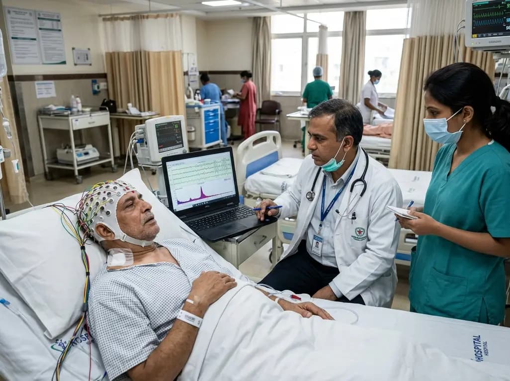
Postoperative delirium represents an acute, fluctuating neurocognitive disturbance that disproportionately affects elderly patients undergoing complex orthopedic and spinal procedures. This clinical syndrome correlates with prolonged hospitalization, increased perioperative morbidity, elevated healthcare costs, and lasting cognitive decline. Although clinicians actively seek non-invasive diagnostic modalities, conventional electroencephalography often fails to capture discrete neurophysiological shifts. Research into postoperative delirium EEG biomarkers has therefore garnered considerable momentum across anesthesiology, neurosurgery, and geriatric care.
Traditional quantitative EEG protocols rely on broad frequency bands, including classic delta, theta, alpha, and beta oscillations. However, these broad spectral averages frequently mask subtle, frequency-specific electrophysiological alterations occurring during acute encephalopathic episodes. Clinicians require refined electrophysiological metrics to accurately detect delirium in its nascent stages. A pioneering investigation published in PLoS One by Lin and colleagues evaluated high-resolution 1-Hz spectral dynamics against traditional broadband analyses in spinal surgery patients. Their exploratory findings demonstrate that granular spectral profiling offers substantially superior diagnostic accuracy compared to conventional broadband EEG assessments.
To systematically evaluate electrophysiological changes, investigators recorded single-electrode frontal EEG from forty-seven older adults undergoing elective spinal surgery. The researchers captured frontal signals using the Fp1 electrode montage across four critical perioperative timepoints. Rather than relying solely on coarse aggregate frequency bands, the analytical framework employed high-resolution 1-Hz spectral bins spanning from 1 Hz to 45 Hz.
Furthermore, the team implemented non-parametric statistical testing coupled with false discovery rate correction to avoid spurious associations. The investigators subsequently assessed diagnostic discrimination using area under the receiver operating characteristic curve metrics. Among the forty-seven enrolled surgical candidates, seven patients developed clinically confirmed delirium, representing an incidence rate of 14.9 percent. This rate closely mirrors the epidemiological incidence observed across broader geriatric surgical cohorts. Consequently, comparing the delirious and non-delirious sub-cohorts yielded profound insights into localized oscillatory disruption. High-resolution single-electrode recording proved straightforward to implement in standard postoperative care units.
The study highlighted a stark performance gap between conventional broadband metrics and high-resolution spectral analyses. Traditional frequency band decomposition revealed only a modest reduction in theta power during active delirium episodes. However, this broadband theta alteration achieved an area under the receiver operating characteristic curve of merely 0.53, indicating virtually no clinical discriminative power.
In sharp contrast, high-resolution 1-Hz spectral analysis revealed a remarkably rich and nuanced neurophysiological landscape. Delirious patients demonstrated significantly elevated 1-Hz spectral power, pronounced power reductions between 3 Hz and 7 Hz, and specific power elevations at 14 Hz and 15 Hz. Most notably, a discrete 23-Hz narrow-band feature achieved the strongest discriminative capability, yielding an area under the curve of 0.74. Interestingly, conventional analysis of the entire beta band (12–30 Hz) failed to reach statistical significance. This discrepancy confirms that broadband averaging dilutes focal oscillatory changes, effectively canceling out meaningful signals within specific narrow frequencies.
A particularly intriguing finding from the investigation involves the temporal persistence of discrete spectral aberrations. Even after overt clinical delirium symptoms subsided according to validated cognitive assessments, several frequency-specific anomalies persisted. Specifically, patients continued to display altered power spectra across narrow frequency channels well into their late postoperative recovery periods.
This sustained electrophysiological disruption suggests that macroscopic behavioral recovery does not immediately mirror full neurobiological normalization. In fact, covert cerebral dysfunction may persist long after patients regain baseline orientation and conversational fluency. Therefore, single-channel frontal EEG biomarkers could serve as objective indicators of ongoing neuronal vulnerability. Clinicians frequently encounter subtle post-discharge cognitive decline in older surgical patients who experienced transient delirium episodes. These persistent spectral abnormalities provide a plausible neurophysiological foundation for prolonged cognitive vulnerability, highlighting the clinical necessity for extended neurocognitive monitoring.
Spine surgery in geriatric populations involves unique physiological stressors, including substantial blood loss, sustained intraoperative positioning challenges, and intensive postoperative analgesic regimens. These compounding factors substantially elevate delirium risk in vulnerable older patients. Implementing point-of-care neuro-monitoring could therefore transform perioperative management across orthopedic and surgical wards.
Moreover, single-electrode frontal recording significantly reduces technical complexity compared with full-montage diagnostic electroencephalography. Anesthesiologists and bedside nursing teams can easily apply frontal adhesive sensors in high-turnover recovery areas. However, commercial depth-of-anesthesia monitors currently utilize coarse proprietary band algorithms that lack granular spectral resolution. Updating monitoring software to include 1-Hz narrow-band parsing could enable real-time detection of delirium-associated neuro-oscillatory signatures. Clinicians could then proactively address reversible triggers, such as electrolyte imbalances, hypoxia, or opioid-induced sedation, before florid behavioral disruptions emerge.
Although these findings offer an exciting diagnostic paradigm, translating high-resolution spectral biomarkers into routine hospital workflows requires careful validation. The exploratory cohort evaluated by Lin and colleagues was relatively small, with seven active delirium cases. Additionally, investigators derived and evaluated diagnostic cutoffs within the same patient cohort, highlighting the need for external validation across diverse multi-center cohorts.
Furthermore, clinicians must distinguish true neurogenic signals from ocular and myogenic artifacts that commonly contaminate frontal Fp1 recordings. High-frequency bands, including the 23-Hz feature, are notoriously susceptible to frontalis muscle tension and micro-saccadic eye movements. Advanced artifact-rejection algorithms and robust signal filtering will prove vital for autonomous bedside devices. Future prospective trials must evaluate whether automated 1-Hz spectral telemetry improves clinical endpoints, such as length of stay, discharge destination, and long-term functional recovery.
Traditional EEG averages electrical activity across broad frequency bands, which dilutes and obscures discrete oscillatory shifts. High-resolution 1-Hz analysis isolates distinct narrow frequencies, such as 23-Hz oscillations, providing significantly greater diagnostic accuracy and revealing subtle neurophysiological disruptions that standard broadband averaging completely overlooks.
Geriatric spine surgery entails extensive surgical trauma, prolonged anesthesia, blood loss, and intense pain management. These physiological stressors significantly increase delirium risk in elderly patients. Delirium correlates with prolonged hospital stays, increased functional decline, higher healthcare costs, and elevated risks of persistent neurocognitive impairment.
Yes, single-electrode frontal EEG from the Fp1 position successfully captures clinically relevant spectral changes. By applying granular 1-Hz spectral analysis, clinicians can detect narrow-band power alterations without requiring cumbersome multi-channel montages, making rapid point-of-care neurocognitive monitoring practical in standard surgical recovery units.
Disclaimer: This content is for informational and educational purposes only. It is not intended to be a substitute for professional medical advice, diagnosis, or treatment. Always seek the advice of your physician or another qualified health provider with any questions you may have regarding a medical condition. Refer to the latest local and national guidelines for clinical practice.
References
Lin G et al. EEG spectral biomarkers of postoperative delirium in spinal surgery: A high-resolution analysis. PLoS One. 2026. doi: 10.1371/journal.pone.0352607. PMID: 42531226.
Bruzzone S et al. Perioperative electroencephalography in predicting and diagnosing postoperative delirium: A systematic review. J Clin Anesth. 2025;98:111634.
Pollak M et al. Electroencephalogram biomarkers from anesthesia induction to identify vulnerable patients at risk for postoperative delirium. Anesthesiology. 2024;140(5):979-989.
Kim J et al. Resting-state prefrontal EEG biomarker in correlation with postoperative delirium in elderly patients. Front Aging Neurosci. 2023;15:1248035.

Read summarized clinical updates, watch expert medical content, and earn CME certifications right from your smartphone.
Open your smartphone camera to scan and install.





A clinical study reveals that 1-Hz high-resolution frontal EEG analysis significantly outperforms conventional broadband EEG in detecting postoperative delirium in spine surgery patients, pinpointing narrow-band biomarkers such as 23-Hz oscillations for enhanced perioperative neuro-monitoring.
last month

Chronic eosinophilic pneumonia can present with atypical bronchoalveolar lavage findings and an organizing pneumonia pattern, necessitating surgical lung biopsy.
Today

Discover the crucial distinctions between mesonephric and mesonephric-like proliferations of the female genital tract. Learn the key histomorphologic, immunohistochemical, and molecular differences essential for accurate diagnosis and clinical management.
Today
Kikuchi-Fujimoto disease rarely presents with refractory shock and sinus bradycardia in children. This case report highlights life-threatening hemodynamic instability requiring intensive care, preserved cardiac contractility, and the critical diagnostic role of early excisional lymph node biopsy.
Today

A narrative review investigates whether the articularis genus muscle functions as an independent anatomical entity or blends with the vastus intermedius. We evaluate its morphology, role in retracting the suprapatellar bursa, and direct clinical significance in anterior knee pain and arthroplasty.
Today

A Bayesian multilevel meta-analysis reveals that aerobic training combined with moderate carbohydrate restriction modestly lowers HbA1c in type 2 diabetes. However, sparse data and very low certainty leave incremental benefits over exercise or diet alone unproven, highlighting the need for individualized care.
Today
