.webp&w=384&q=75)

Loading, please wait...
Loading, please wait...

The posterolateral corner of the knee remains one of the most structurally intricate anatomical regions encountered in sports orthopedics. Mastering popliteal hiatus anatomy is critical for clinicians evaluating lateral knee pain, meniscal extrusion, and posterosuperior instability. Unlike the medial meniscus, which adheres firmly along the joint capsule, the lateral meniscus lacks a continuous peripheral attachment where the popliteus tendon traverses the joint. Instead, dynamic ligamentous bands and fascial slips tether the meniscus, preventing excessive translation during rotational stress. Recent anatomical and histological investigations have revealed surprising structural characteristics of these soft tissues. Consequently, these findings reshape how arthroscopic surgeons understand lateral meniscal stability and guide biologically augmented surgical repair strategies.
The popliteus hiatus represents a distinct physiological defect in the coronary ligament through which the popliteus musculotendinous unit travels. Because the lateral meniscus lacks direct peripheral bone anchors here, secondary stabilizers must preserve lateral joint mechanics. Historically, surgeons viewed the capsular boundaries surrounding the popliteus hiatus primarily as thin membranous folds. However, modern kinematic biomechanical analyses show that disruptions here create hypermobility of the lateral meniscus. Consequently, patients present with unexplained lateral joint-line tenderness, painful catching sensations, or sudden locking episodes during deep squatting movements.
Furthermore, dynamic loading during knee flexion induces significant posterior translation of the lateral meniscus. The popliteomeniscal complex restrains this motion while permitting necessary physiological excursion. Therefore, when these supporting bands suffer traumatic rupture or microtears, lateral meniscal hypermobility ensues. This subtle pathology frequently accompanies acute anterior cruciate ligament tears or occurs as an isolated athletic injury. Orthopedic specialists often overlook these hidden hiatal disruptions during initial evaluations because magnetic resonance imaging frequently appears normal. As a result, comprehensive familiarity with this anatomical crossroad enables surgeons to recognize rotational laxity and avoid erroneous partial meniscectomies that worsen joint contact pressures.
Detailed cadaveric dissections have mapped the intricate relationship among the popliteomeniscal fascicles, the popliteofibular ligament, and the meniscofibular ligament. Specifically, researchers dissected fresh-frozen knee specimens to examine these distinct fibrous structures. The posterosuperior popliteomeniscal fascicle originates consistently from the posterior aspect of the popliteus tendon and inserts onto the superior margin of the lateral meniscus. In contrast, the posteroinferior popliteomeniscal fascicle originates from the anterior border of the popliteus tendon before inserting onto the inferior meniscal rim.
Importantly, dense adipose tissue reliably separates the posterosuperior and posteroinferior fascicles, creating a combined encapsulation around the traversing popliteus tendon. In addition, an interfascicular interval cleanly separates the posteroinferior fascicle from the meniscofibular ligament in most knees. The meniscofibular ligament arises directly from the inferior rim of the lateral meniscus, connects superiorly with the anterior popliteomeniscal fascicle, and converges distally with the popliteofibular ligament at the fibular head. Thus, these structures do not represent haphazard synovial reflections. Instead, they form an organized three-dimensional suspension apparatus that stabilizes both the meniscus and the posterolateral corner during complex multi-planar rotational movements.
Histological evaluation provides critical insights into the biological composition and structural strength of these peri-hiatal tissues. Recent histological analyses demonstrate marked microscopic differences between the individual popliteomeniscal components. Specifically, staining reveals that the meniscofibular ligament and the posteroinferior popliteomeniscal fascicle possess true ligamentous architecture. They feature parallel arrays of densely packed collagen bundles interspersed with elongated, spindle-shaped fibroblasts. These structural characteristics confirm their direct role in bearing mechanical tensile loads during knee flexion and external tibial rotation.
Conversely, histological examination of the anterior popliteomeniscal fascicle and posterosuperior popliteomeniscal fascicle reveals a strikingly different histological profile. Rather than dense collagenous fascicles, these components display vascularized synovial morphology with loose fibrovascular stroma. Therefore, the posterosuperior fascicle and anterior fascicle act primarily as protective, vascularized synovial conduits rather than rigid tensile restraints. This distinction fundamentally alters historical assumptions that treated all fascicles as identical mechanical suspensors. Recognizing that the posteroinferior fascicle and meniscofibular ligament represent true ligaments underscores their indispensable role in maintaining meniscal stability. Moreover, this biological insight explains why tearing these specific ligamentous structures causes symptomatic joint instability.
Diagnosing lesions within the popliteal hiatus area remains exceptionally difficult in routine clinical practice. Standard knee magnetic resonance imaging frequently fails to delineate isolated fascicular injuries due to oblique orientations and partial volume averaging. Consequently, clinicians must maintain high suspicion when athletes report persistent lateral clicking, localized discomfort, or recurrent effusion without frank meniscal body tears. Clinical tests, including the figure-four test and forced external rotation in deep flexion, may reproduce symptoms.
Furthermore, diagnostic arthroscopy requires systematic inspection of the posterolateral hiatus beyond standard anterior portal visualization. Surgeons frequently employ the arthroscopic drive-through sign or probe-based aspiration testing to assess excessive mobility. Under normal conditions, a surgeon cannot displace the posterior horn of the lateral meniscus into the intercondylar notch. However, tears involving the posteroinferior fascicle or meniscofibular ligament permit abnormal anterior translation greater than five millimeters. In addition, surgeons must carefully evaluate the lateral gutter to confirm whether the popliteus tendon separates abnormally from the meniscal border. By methodically probing the popliteomeniscal boundaries, arthroscopists can detect occult structural laxity before cartilage wear develops.
Because the meniscofibular ligament and posteroinferior popliteomeniscal fascicle consist of dense, hypovascular ligamentous tissue, intrinsic healing capacity remains intrinsically poor after injury. Conservative management or simple debridement frequently fails to relieve mechanical locking and persistent pain. Therefore, modern sports surgeons advocate for anatomical arthroscopic repair to restore lateral compartment kinematics. Clinicians can utilize an all-inside suture hook technique or modern low-profile meniscal repair devices to re-anchor the torn ligamentous fascicles directly back to the popliteus sheath and joint capsule.
Moreover, biological augmentation plays a pivotal role in ensuring solid fibrocartilaginous healing across the repair site. Surgeons strongly recommend combining suture stabilization with mechanical trephination or microfracture of the adjacent popliteus tendon and vascular bed. Trephination stimulates local bleeding and releases autologous growth factors that promote collagen synthesis within the hypovascular zone. In addition, some surgeons introduce concentrated bone marrow aspirate or platelet-rich plasma to optimize cellular regeneration. Restoring structural continuity protects the knee from abnormal shear forces and slows premature osteoarthritis progression. Ultimately, matching meticulous mechanical fixation with targeted biological enhancement ensures optimal patient recovery and sustained athletic performance.
The popliteomeniscal fascicles are three distinct fibrous bands located around the popliteal hiatus of the lateral meniscus. They comprise the posterosuperior, posteroinferior, and anterior fascicles. These structures bridge the gap where the lateral meniscus lacks standard capsular attachment around the traversing popliteus tendon. Consequently, they anchor the lateral meniscus, prevent meniscal entrapment during joint motion, and maintain essential posterolateral knee stability throughout varied degrees of flexion and tibial rotation.
Histological composition directly influences the intrinsic healing potential of musculoskeletal tissues. Because the meniscofibular ligament and posteroinferior popliteomeniscal fascicle possess dense, hypovascular ligamentous architecture, they cannot heal spontaneously after complete tears. Conversely, the vascular synovial nature of the posterosuperior fascicle supports intrinsic healing. Therefore, recognizing true ligamentous tissue prompts orthopedic surgeons to perform mechanical suture fixation alongside biological augmentation, such as trephination or microfracture, to stimulate blood flow and facilitate robust tissue regeneration.
Patients suffering from a hypermobile lateral meniscus typically report persistent lateral joint pain, mechanical catching, or locking during deep knee flexion and squatting. Furthermore, standard magnetic resonance scans often miss these injuries because static images show intact meniscal fibrocartilage. Clinicians frequently confirm instability during arthroscopy by performing the drive-through test or probe manipulation. When stabilizing fascicles are disrupted, the posterior horn translates abnormally beyond five millimeters into the joint, confirming significant mechanical compromise.
Disclaimer: This content is for informational and educational purposes only... Refer to the latest local and national guidelines for clinical practice.
References

Read summarized clinical updates, watch expert medical content, and earn CME certifications right from your smartphone.
Open your smartphone camera to scan and install.





New anatomical and histological evidence reveals that the meniscofibular ligament and posteroinferior popliteomeniscal fascicle are true ligamentous structures. Discover the diagnostic and surgical repair implications for posterolateral knee instability.
Today
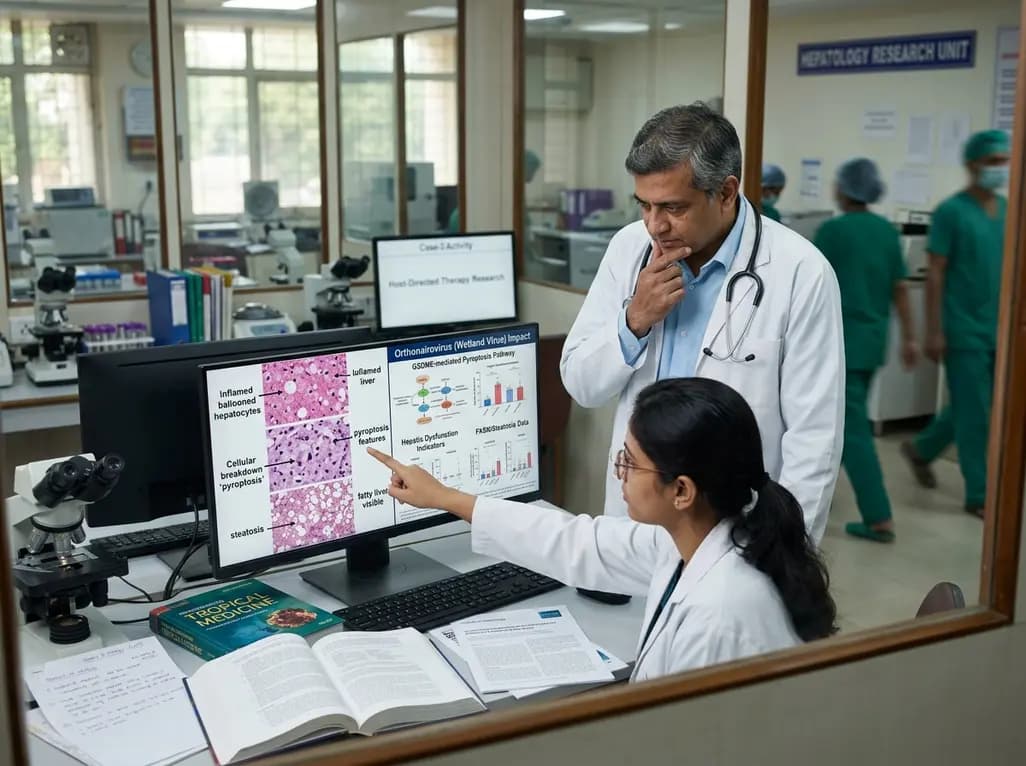
Newly recognized orthonairoviruses like Wetland virus trigger lethal hepatic dysfunction via caspase-3-gasdermin E pyroptosis and fatty acid synthase-dependent steatosis. This crucial link between inflammatory cell death and metabolic dysregulation highlights novel avenues for host-directed antiviral therapies.
Today

A cross-sectional study of 700 college students highlights significant contraceptive knowledge disparities between sexually active and inactive young adults. The findings demonstrate a critical need for proactive, comprehensive sexual health counseling before sexual debut.
Today
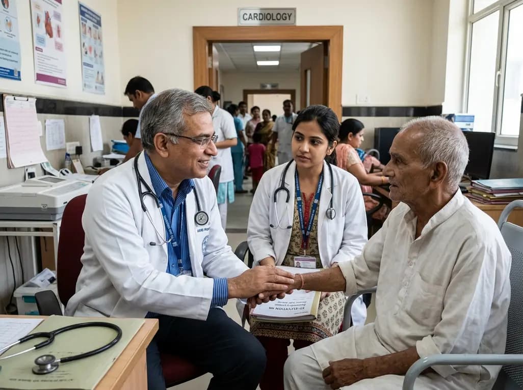
A landmark nationwide cohort study reveals that older adults with dementia who undergo cardiac catheterization for STEMI spend meaningful time alive at home, particularly community-dwelling individuals. Cognitive impairment alone should not preclude acute invasive revascularization.
Today

A randomized controlled trial demonstrates that theory-driven personalized mobile messaging reduces the risk of consecutive walking goal failures by 33 percent, highlighting the vital role of digital health tools in sustaining long-term exercise habits and chronic disease prevention.
Today
