.webp&w=384&q=75)

Loading, please wait...
Loading, please wait...
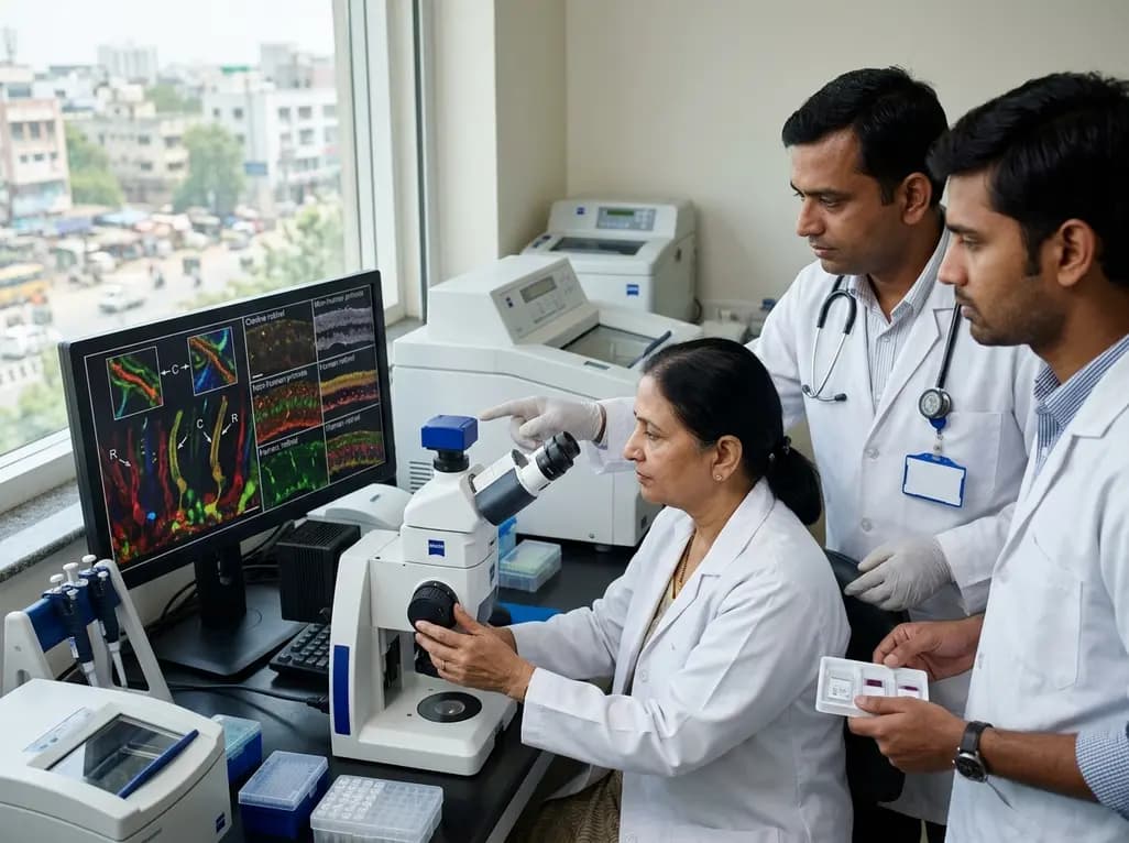
The vertebrate retina relies on highly specialized sensory neurons to capture light and initiate vision. At the center of this phototransduction process lies the photoreceptor sensory cilium, a modified primary cilium that bridges the metabolically active inner segment with the light-sensitive outer segment. Photoreceptors continuously synthesize opsins and auxiliary signaling molecules within the inner segment, which must traffic across this narrow ciliary conduit at high rates. Consequently, any structural defect or molecular disruption within this sensory apparatus can lead to severe inherited retinal dystrophies. Recent advancements in ultrastructure expansion microscopy have allowed researchers to visualize these micro-domains with unprecedented nanoscale resolution. By comparing canine, non-human primate, and human retinas, investigators have successfully mapped the complex molecular organization of both ciliary and periciliary compartments. These comparative findings illuminate how fundamental structural archetypes remain conserved across evolutionary lineages while distinct morphological specializations adapt to specific species needs. Ultimately, understanding these nanoscale differences provides clinicians and ophthalmic researchers with crucial insights into the mechanisms underlying retinal ciliopathies, disease progression, and prospective translational therapeutics.
The core structural framework of the photoreceptor sensory cilium demonstrates remarkable evolutionary conservation among large mammals. Ultrastructural investigations show that the microtubule-based ciliary axoneme extends from the basal body and traverses the connecting cilium before reaching into the phototransductive outer segment. Furthermore, this structural continuum maintains the physical integrity of the entire outer retinal layer. Researchers observed that the transition zone machinery, essential for gating protein transport, exhibits uniform spatial distribution across canines, non-human primates, and humans. In addition, key scaffolding elements and structural microtubule doublets align precisely along the longitudinal axis of the ciliary shaft. Therefore, fundamental transport processes, including intraflagellar transport mechanisms, follow identical biophysical rules in disparate mammalian eyes. This high level of structural conservation emphasizes the indispensable role of the ciliary backbone in supporting outer segment disk morphogenesis. Moreover, these shared molecular baselines explain why fundamental phototransduction cascades function similarly across mammalian species, providing a solid foundation for cross-species comparative biology.
Although the fundamental framework remains conserved, significant architectural differences distinguish rod photoreceptors from cone photoreceptors across all examined species. Cones systematically display shorter connecting cilia compared to rods, reflecting differences in their metabolic turnover and inner segment proximity. Additionally, cone photoreceptors feature markedly longer daughter centrioles and an enlarged ciliary bulge region at the proximal outer segment junction. In contrast, rods possess a more slender, elongated connecting cilium that supports continuous, unidirectional disk displacement toward the retinal pigment epithelium. Cone outer segments maintain open-surface lamellae that require distinct periciliary anchoring compared to the closed, internalized disks characteristic of rod outer segments. Consequently, these structural variations modulate the kinetic rates of protein trafficking between cell types. These intrinsic structural differences also explain why certain genetic mutations preferentially compromise rod function, leading to retinitis pigmentosa, whereas other variants selectively trigger cone-rod dystrophies or macular degenerations in ophthalmic practice.
Beyond the central ciliary axoneme, periciliary structures exhibit substantial species-specific diversification, particularly in rod photoreceptors. Non-human primate and human retinas demonstrate circumferential periciliary membranes paired with robust, well-developed calyceal processes that embrace the base of the outer segment. In stark contrast, canine rods display a unilateral periciliary membrane configuration and completely lack classical calyceal processes. However, cone photoreceptors across all three species maintain prominent calyceal processes, suggesting an essential mechanical role in stabilizing cone outer segments. Furthermore, human photoreceptors exhibit notable inter-individual variability in accessory inner segment-like structures that are less prominent in non-human primates. These species-dependent variations highlight how evolutionary pressures have modified accessory structural supports to accommodate differences in eye size, mechanical forces, and diurnal activity patterns. Consequently, recognizing these anatomical distinctions prevents erroneous generalizations when translating mechanistic data from animal models directly to human retinal physiology.
Retinal ciliopathies encompass a heterogeneous group of genetic disorders characterized by progressive visual impairment and photoreceptor degeneration. Prominent examples include Usher syndrome, Bardet-Biedl syndrome, and Leber congenital amaurosis, where ciliary transport breakdown precipitates outer segment atrophy. Because canines naturally develop inherited retinal dystrophies homologous to human conditions, they serve as invaluable translational models for pre-clinical studies. Nevertheless, structural disparities in periciliary architecture mandate caution when interpreting rescue phenotypes and transport kinetics. For example, mutations affecting calyceal process-associated proteins, such as standard Usher syndrome complexes, may exhibit divergent cellular pathology in species lacking these specific supports in rods. Therefore, detailed nanoscale molecular maps enable clinicians and scientists to correlate specific clinical phenotypes with precise structural deficits. Additionally, these cross-species comparisons help researchers predict whether therapeutic responses observed in large animal models will accurately reflect human treatment outcomes during clinical trials.
The refinement of ciliary structural biology directly enhances diagnostic accuracy and therapeutic design in modern ophthalmic genetics. Advanced genetic testing frequently identifies novel variants of uncertain significance within ciliary structural genes. Clinicians can now contextualize these variants against high-resolution molecular maps of the transition zone, basal foot, and periciliary membrane domains. Furthermore, novel ocular gene delivery platforms, such as adeno-associated viral vectors and lipid nanoparticles, require precise targeting to photoreceptor compartments. Understanding the exact nanoscale dimensions and physical constraints of the connecting cilium allows bioengineers to optimize capsid design and promoter specificity. In addition, evaluating individual anatomical variations reinforces the necessity for personalized therapeutic windows before irreversible photoreceptor cell loss occurs. Ultimately, bridging molecular structural findings with clinical genetics equips ophthalmologists to offer more accurate prognostic counseling and tailored therapeutic interventions for patients with blinding inherited retinal diseases.
The primary function involves facilitating bidirectional molecular transport between the inner and outer segments of retinal photoreceptors. Synthesized phototransduction proteins, such as rhodopsin, travel through this narrow ciliary bridge to maintain outer segment structure. Without an intact ciliary transport system, photoreceptor cells rapidly degenerate, causing severe vision loss.
Cone photoreceptors systematically display shorter connecting cilia, longer daughter centrioles, and larger ciliary bulges than rods across mammalian species. In addition, cones feature open outer segment lamellae supported by prominent calyceal processes, whereas rods possess closed internal disks with species-variable periciliary membranes.
Cross-species variations determine how faithfully animal models recapitulate human retinal disease mechanisms. Because canine rods lack calyceal processes present in primates, therapeutic responses to structural gene delivery may differ between species. Recognizing these variations ensures safer translation and better vector optimization for human clinical trials.
Disclaimer: This content is for informational and educational purposes only and should not be considered medical advice. Always consult a qualified healthcare professional regarding any medical condition or treatment. Refer to the latest local and national guidelines for clinical practice.
References
1. Takahashi K et al. Cross-species molecular mapping of the photoreceptor sensory cilium and periciliary structures identifies conserved and species-specific architectural features. J Cell Sci. 2026 Sep 01. doi: undefined. PMID: 42676275.
2. Takahashi K, Obayashi M, Sudharsan R, Beltran WA. Mapping Protein Distribution in the Canine Photoreceptor Sensory Cilium and Calyceal Processes by Ultrastructure Expansion Microscopy. Invest Ophthalmol Vis Sci. 2025;66(2):1.
3. Bachmann-Gagescu R, Neuhauss SCF. The photoreceptor cilium and its diseases. Curr Opin Genet Dev. 2019;56:22-33.

Read summarized clinical updates, watch expert medical content, and earn CME certifications right from your smartphone.
Open your smartphone camera to scan and install.





Nanoscale molecular mapping using expansion microscopy reveals conserved rod-cone structural divergence and species-specific variations in the photoreceptor sensory cilium across canine, non-human primate, and human retinas, offering critical insights for inherited retinal diseases and translational gene therapies.
4 weeks back
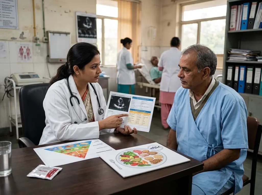
A scientific statement highlights the crucial role of nutrition in managing chronic heart failure. It emphasizes GLIM-based screening, individualized dietary patterns like the Mediterranean diet, appropriate protein and energy intake, avoiding unnecessary fluid restriction, and targeted iron supplementation.
Today

A nationwide South Korean study shows underreporting of smoking among women dropped from 57.4% to 36.4% between 2008 and 2021. However, over one-third of female smokers still conceal their habit, highlighting the critical role of cotinine verification in clinical risk assessment.
Today

PepsiCo and Monster Beverage have challenged the FSSAI ban on the energy drink label in Indian courts. While companies highlight supply disruptions, regulatory actions spotlight severe cardiovascular and metabolic risks associated with excessive caffeine, sugar, and taurine consumption, especially in adolescents.
Today

A new study evaluates the rheological stability of decellularized peripheral nerve matrix hydrogels under varied storage conditions. Findings show -80°C storage and freeze-drying preserve gelation kinetics and mechanical stiffness, offering key insights for shipping and translational clinical applications.
Today

A qualitative meta-synthesis highlights the complex interplay between gender dysphoria, minority stress, and eating disorders in transgender and gender-diverse individuals. Clinicians must recognize these distinct drivers to offer supportive, gender-affirming, and interdisciplinary eating disorder care.
Today
