.webp&w=384&q=75)

Loading, please wait...
Loading, please wait...

Perinatal intracranial haemorrhage in full-term infants represents a grave clinical challenge requiring urgent multidisciplinary intervention. Unlike preterm hemorrhage, bleeding in term infants often stems from distinct mechanical, vascular, or coagulopathic etiologies. Understanding developmental trajectories and objective prognostic markers is essential for pediatricians and neurologists. Recent prospective investigations provide crucial evidence regarding the long-term neurodevelopmental consequences and structural predictors of functional disability.
Perinatal intracranial bleeding in term neonates exhibits marked clinical heterogeneity. In a landmark prospective study of sixty-eight term infants, researchers evaluated children longitudinally from the perinatal window into childhood. Interestingly, approximately two-thirds of the cohort received a diagnosis postnatally, whereas one-third was identified antenatally during obstetrical ultrasonography.
Furthermore, intraventricular haemorrhage emerged as the most prevalent bleeding phenotype, occurring in over sixty-three percent of all subjects. Notably, intraventricular bleeding occurred significantly more often in fetuses diagnosed prenatally than in neonates diagnosed after birth. In contrast to premature infants whose bleeding typically originates in the fragile subependymal germinal matrix, term infants frequently exhibit multi-compartment bleeding involving subdural, subarachnoid, and parenchymal regions. Delivery trauma, perinatal asphyxia, sinovenous thrombosis, and coagulopathies frequently contribute to these presentations. Consequently, identifying the exact anatomic distribution during initial neuroimaging remains critical for establishing baseline clinical severity and guiding timely neonatology management. Moreover, prompt identification of underlying hemostatic defects prevents ongoing intracranial rebleeding.
Longitudinal evaluation reveals that intracranial bleeding in term infants conveys substantial risk for persistent neurological disability. Researchers assessed functional outcomes at a median age of nearly four years using the Pediatric Stroke Outcome Measure and the modified Rankin Scale. Strikingly, almost forty-three percent of the cohort received a formal diagnosis of cerebral palsy during childhood surveillance.
Additionally, functional outcomes varied widely across the study population. Normal neurodevelopment occurred in only forty-two percent of patients. Conversely, nearly eighteen percent displayed mild disability, sixteen percent exhibited moderate impairment, and over twenty-three percent suffered severe neurological deficits. Crucially, researchers discovered no significant difference in neurological prognosis between infants diagnosed antenatally and those identified postnatally. This vital finding demonstrates that prenatal identification neither assures milder disease nor dictates an inherently worse prognosis. Instead, total tissue disruption and structural parenchymal damage determine functional destiny. Pediatricians must therefore counsel families objectively, avoiding assumptions based solely on the chronological timing of initial radiological discovery. Furthermore, ongoing parent education regarding early developmental milestones ensures rapid identification of delayed motor skills.
Advanced magnetic resonance imaging serves as the cornerstone for predicting functional impairment after perinatal bleeding. In the prospective study, specific anatomical lesions demonstrated statistically significant correlations with unfavourable stroke outcome scores. Multi-compartment bleeding, cerebellar vermian haemorrhage, and posthaemorrhagic ventricular dilatation strongly heralded compromised motor and cognitive performance.
Furthermore, parenchymal damage patterns yielded profound prognostic value. Significant thalamic volume loss, extensive cerebral white matter reduction, and Wallerian degeneration within descending corticospinal tracts directly correlated with poorer developmental metrics. Similarly, abnormal myelination in the posterior limb of the internal capsule served as a robust radiologic hallmark of future cerebral palsy. Radiologists also identified a significant correlation between anterior horn width and cumulative disability scores. When ventricular dilatation expands the anterior horn, secondary pressure leads to periventricular axonal injury. Consequently, systematic MRI scoring allows clinicians to identify high-risk infants before clinical deficits fully manifest. Clinicians can then implement preemptive neuroprotective strategies and initiate early structured therapy.
Remote epilepsy represents one of the most debilitating long-term complications of perinatal brain damage. Within the studied cohort, over nineteen percent of term infants developed chronic epilepsy. Importantly, multivariable statistical analysis confirmed a strong association between remote unprovoked seizures and severely compromised neurodevelopmental scores.
Pathophysiologically, cortical blood extravasation promotes hemosiderin deposition, recurrent microvascular ischemia, and extensive reactive astrogliosis. These structural alterations generate persistent epileptogenic networks within neonatal cerebral tissue. Moreover, recurring unprovoked seizures disrupt early synaptogenesis and impair neuroplasticity during formative neurodevelopmental windows. This secondary epileptic encephalopathy aggravates motor deficits and accelerates cognitive regression. Therefore, pediatric neurologists must maintain vigilant surveillance for post-neonatal seizures. Routine electroencephalography monitoring helps identify subclinical epileptiform discharges before generalized seizures emerge. Additionally, routine screening for subclinical visual and motor auras helps clinicians optimize pharmacotherapy before secondary neurological injury expands. Decisive antiseizure drug therapy, combined with parental education on seizure semiology, remains essential for protecting developing cognitive networks and preventing further neurological deterioration.
Given that over fifty percent of surviving term infants with intracranial hemorrhage experience mild to severe neurodevelopmental deficits, proactive clinical management is mandatory. Following acute neonatal stabilization, pediatric clinicians should enroll infants into comprehensive early intervention programs. An integrated multidisciplinary team comprising pediatric neurologists, physical therapists, occupational therapists, and speech specialists provides customized rehabilitative support.
Specifically, focused physiotherapy prevents spastic contractures, while occupational therapy fosters bilateral motor coordination and adaptive functional play. Speech and language interventions mitigate communication delays resulting from cortical and subcortical injury. In Indian healthcare systems, early enrollment in district intervention facilities ensures sustained rehabilitation access across diverse socioeconomic backgrounds. Longitudinal follow-up must continue through school age, utilizing standardized testing scales to evaluate emerging cognitive, executive, and behavioral challenges. Additionally, serial cranial ultrasound and magnetic resonance imaging track ventriculomegaly, guiding timely neurosurgical referral when posthaemorrhagic hydrocephalus threatens brain parenchyma. Ultimately, early identification and continuous multidisciplinary surveillance dramatically improve functional outcomes, empowering affected children to achieve their highest developmental potential.
Prospective evidence indicates that the timing of diagnosis does not significantly influence long-term neurological outcomes. Full-term infants diagnosed antenatally demonstrate neurodevelopmental disability rates comparable to those diagnosed postnatally. Instead, outcome severity depends directly on underlying anatomical parenchymal involvement, secondary ventriculomegaly, thalamic damage, and associated remote epileptic activity.
Key magnetic resonance imaging markers predicting poor outcomes include multi-compartment hemorrhage, cerebellar vermian involvement, and posthaemorrhagic ventricular dilatation. In addition, thalamic volume reduction, supratentorial white matter loss, Wallerian degeneration along the corticospinal tracts, and abnormal myelination within the posterior limb of the internal capsule strongly correlate with adverse developmental scores.
Early rehabilitation capitalizes on maximal infant neuroplasticity before pathological motor patterns and joint contractures become established. Multidisciplinary interventions combining physical, occupational, and speech therapy significantly improve functional motor trajectories, optimize sensory processing, prevent secondary musculoskeletal deformities, and enhance long-term independence in children experiencing cerebral palsy or cognitive impairment.
Disclaimer: This content is for informational and educational purposes only. It is not intended to provide medical advice or to be a substitute for professional medical advice, diagnosis, or treatment. Patients should always seek the advice of their physician or other qualified healthcare provider regarding a medical condition. Clinicians should rely on their clinical judgment and check appropriate medical resources before initiating or changing treatments. Refer to the latest local and national guidelines for clinical practice.
References

Read summarized clinical updates, watch expert medical content, and earn CME certifications right from your smartphone.
Open your smartphone camera to scan and install.





Prospective research shows that term infants with perinatal intracranial haemorrhage face significant long-term risks of cerebral palsy and remote epilepsy. Advanced MRI markers, such as thalamic volume loss and ventriculomegaly, provide essential prognostic guidance for early multidisciplinary intervention.
Today

A multicity study highlights that over 80% of acute respiratory infections remain outside hospitals. Patients frequently choose community pharmacies and digital platforms. Integrating digital health data and retail pharmacy sales can fill critical surveillance gaps and improve early outbreak detection.
Today

Scandinavian register evidence reveals that neonatal birth length strongly predicts adult health and mortality, whereas birth weight dictates cognitive capacity. Symmetric growth restriction causes far more severe long-term deficits than asymmetric restriction, underscoring the need for comprehensive anthropometry.
Today
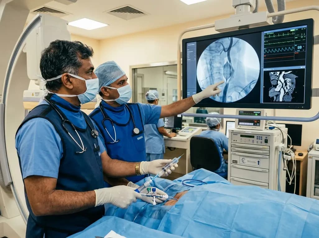
Discover how rescue cutting balloon angioplasty successfully modifies heavily calcified carotid stenosis, enabling stent delivery without baroreceptor instability and ensuring favorable long-term neurologic outcomes.
Today
A breakthrough study reveals that digoxin alleviates osteoarthritis by targeting LSP1 in synovial macrophages, preventing harmful extracellular trap formation and restoring SOD1 activity. This dual-action mechanism protects joint cartilage and offers a promising repurposing strategy for disease modification.
Today

Data from the 2026 Workforce Health Index reveals that 38.7% of urban employees and dependents exhibit abnormal glycemic control. Emerging at increasingly younger ages, this metabolic shift intersects with severe vitamin deficiencies, highlighting the urgent need for early workplace screenings and targeted clinical care.
Today
