.webp&w=384&q=75)

Loading, please wait...
Loading, please wait...

Traumatic brain injury remains a primary cause of morbidity and death in children worldwide. Evaluating acute pediatric head trauma poses substantial challenges for clinicians who must identify intracranial hemorrhage while limiting harmful radiation exposure. Fortunately, recent prospective studies have demonstrated the transformative clinical value of pediatric TBI biomarkers in acute neurotrauma evaluation. By quantifying circulating glial and neuronal proteins released into the bloodstream after mechanical impact, clinicians can gain objective insight into central nervous system pathology.
Emergency physicians routinely evaluate children presenting after accidental falls, motor vehicle accidents, and recreational injuries. Determining whether an injured infant or adolescent requires an urgent cranial computed tomography (CT) scan remains challenging. While non-contrast head CT effectively identifies acute traumatic intracranial lesions, medical ionizing radiation carries documented lifetime carcinogenic risks in children. Furthermore, uncooperative or distressed pediatric patients frequently require chemical sedation to complete neuroimaging, introducing additional procedural hazards. Validated clinical decision rules, such as the Pediatric Emergency Care Applied Research Network (PECARN) criteria, reliably identify children at very low risk of clinically important traumatic brain injury. Nevertheless, a substantial intermediate-risk group presents with clinical signs such as persistent vomiting, brief loss of consciousness, or isolated scalp hematomas. Consequently, clinicians often order emergency CT scans out of caution to avoid missing acute intracranial bleeding. As a result, pediatric medicine requires non-invasive, objective biological tools to refine risk stratification and prevent unnecessary neuroimaging.
Recent findings from the pediatric cohort of the Transforming Research and Clinical Knowledge in Traumatic Brain Injury (TRACK-TBI) study provide crucial evidence on molecular diagnostics. Researchers evaluated 70 pediatric trauma patients who had blood sampling and study-reviewed cranial CT or magnetic resonance imaging (MRI). The investigators quantified plasma concentrations of glial fibrillary acidic protein (GFAP), ubiquitin carboxy-terminal hydrolase-L1 (UCH-L1), S100B, and high-sensitivity C-reactive protein (hsCRP). Statistical analyses showed marked differences in day-of-injury biomarker levels between CT-positive and CT-negative patients. Specifically, multivariable linear regression confirmed that plasma GFAP, UCH-L1, and S100B independently associated with acute traumatic findings on head CT. Moreover, GFAP demonstrated a significant association with persistent traumatic intracranial abnormalities detected on two-week follow-up MRI. Among all evaluated proteins, GFAP served as the strongest predictor of neuroimaging findings on receiver operating characteristic analysis. Consequently, these robust data confirm that circulating brain proteins reliably reflect traumatic intracranial damage across diverse pediatric injury severities.
Understanding the cellular origins of these circulating markers explains their diagnostic accuracy in acute pediatric head trauma. Glial fibrillary acidic protein serves as an essential intermediate filament in mature astrocytes. When biomechanical forces disrupt astrocytic cell membranes or compromise the blood-brain barrier, GFAP rapidly leaks into extracellular cerebral fluid and peripheral circulation. In contrast, ubiquitin carboxy-terminal hydrolase-L1 is an abundant enzyme localized within neuronal cell bodies that regulates intracellular protein degradation. Following acute axonal shearing or neuronal necrosis, injured neurons promptly release UCH-L1 into systemic capillaries. Therefore, measuring both proteins simultaneously provides a comprehensive snapshot of glial and neuronal structural integrity. Meanwhile, S100B acts as a calcium-binding protein synthesized primarily by astroglial cells, although extra-cranial tissues like adipocytes and chondrocytes also express minor quantities. In the TRACK-TBI analysis, GFAP significantly outperformed S100B and inflammatory markers like hsCRP in distinguishing intracranial pathology. Thus, astroglial structural proteins offer exceptional specificity for cerebral tissue damage.
Interpreting circulating neurotrauma markers in pediatric patients requires careful consideration of baseline neurodevelopment and time elapsed since injury. Notably, the TRACK-TBI cohort identified significant age-dependent differences for circulating S100B and neuron-specific enolase, showing substantially higher physiological levels in infants and toddlers. Normal synaptogenesis, brain growth, and physiological blood-brain barrier permeability explain these elevated baselines during early childhood. Because adult diagnostic thresholds do not apply directly to young children, establishing age-stratified reference intervals remains essential for clinical practice. In addition to age, clinicians must account for divergent biomarker clearance kinetics. For example, UCH-L1 concentrations peak rapidly within hours of trauma but decline swiftly after eight hours. Conversely, plasma GFAP levels rise gradually and remain detectable for several days post-injury. Therefore, clinicians must document the exact time of injury before interpreting blood test results. Properly timing the blood draw ensures accurate detection of acute parenchymal injury while avoiding false-negative determinations.
Integrating objective blood biomarkers into routine trauma protocols could revolutionize pediatric emergency care worldwide, including in Indian hospitals. In tertiary trauma centers, combining validated decision algorithms like PECARN with rapid point-of-care biomarker testing can decisively resolve intermediate-risk cases. If an ambiguous patient displays normal biomarker concentrations, clinicians can safely avoid cranial CT scans and prioritize clinical observation. Consequently, emergency departments can reduce hospital observation times, prevent radiation-induced malignancies, and spare parents unnecessary emotional distress. Furthermore, in resource-constrained environments where access to emergency CT scanners remains limited, rapid biomarker testing can guide urgent inter-facility transfers. However, successful healthcare implementation requires overcoming cost barriers and establishing automated point-of-care testing platforms in emergency units. Additionally, clinical laboratories must standardize age-adjusted cutoff values across diverse patient demographics. Ultimately, adopting blood-based biomarkers will empower emergency physicians, pediatricians, and neurosurgeons to make objective, evidence-based neuroimaging decisions.
Biomarkers like GFAP and UCH-L1 indicate structural brain injury after trauma. When children present with intermediate-risk head trauma, normal biomarker concentrations reliably indicate the absence of acute intracranial lesions. Consequently, clinicians can safely avoid unnecessary ionizing radiation from head CT scans and shorten emergency observation periods.
Children undergo dynamic neurodevelopment, synaptic pruning, and blood-brain barrier maturation throughout infancy and childhood. Consequently, physiological baseline concentrations of proteins like GFAP, S100B, and NSE are naturally higher in young pediatric cohorts than in adults. Clinicians must apply validated age-stratified cutoffs to prevent false-positive interpretations and unnecessary hospitalizations.
Glial fibrillary acidic protein reflects astroglial cell injury and remains elevated in blood for several days following mechanical trauma. Conversely, ubiquitin carboxy-terminal hydrolase-L1 marks acute neuronal body damage and peaks rapidly within hours before declining. Measuring both proteins provides complementary insights across different cellular structures and injury timeframes.
Disclaimer: This content is for informational and educational purposes only and does not constitute medical advice, diagnosis, or treatment. Healthcare professionals should rely on their clinical judgment, institutional protocols, and current medical evidence when evaluating and managing patients. The findings discussed reflect recent research and may not apply to all clinical scenarios. Refer to the latest local and national guidelines for clinical practice.
References

Read summarized clinical updates, watch expert medical content, and earn CME certifications right from your smartphone.
Open your smartphone camera to scan and install.





A TRACK-TBI study reveals that plasma biomarkers GFAP, UCH-L1, and S100B correlate strongly with acute CT and MRI abnormalities in pediatric head trauma. These blood markers provide objective risk stratification, identifying intracranial injury while reducing unnecessary radiation from cranial CT scans in children.
Today
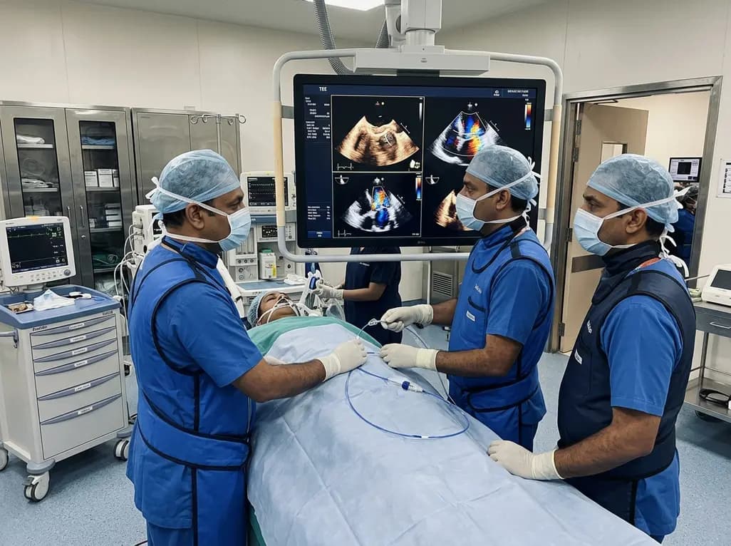
Transcatheter edge-to-edge repair (TEER) successfully treated severe mitral regurgitation from a posterior leaflet perforation. Guided by 3D multiplanar reconstruction, operators sandwiched the defect using two MitraClip devices, providing safe hemodynamic recovery without requiring open-heart surgery.
Today
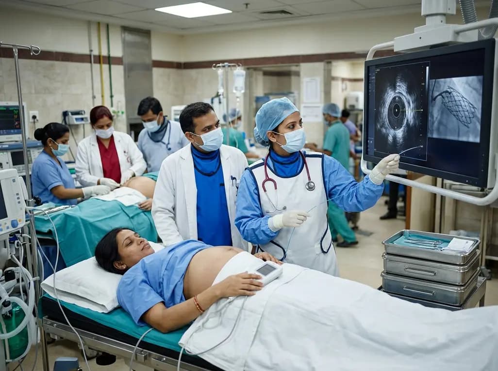
A 32-year-old gravida at 38 weeks with anterior STEMI underwent emergent IVUS-guided PCI and successful cesarean delivery under uninterrupted DAPT, highlighting cardio-obstetric care.
Today

A systematic review and meta-analysis of 5,406 participants reveals that tirzepatide significantly reduces hsCRP by 34.3%, improves cardiorenal biomarkers, and enhances functional capacity and exercise endurance in patients with heart failure.
Today

A landmark biomechanical study demonstrates that sports bra support and running shoe cushioning interact significantly to influence tibial acceleration, breast motion, and comfort in female runners, providing crucial insights for clinical counseling in sports medicine and women's health.
Yesterday

New ADIPO-CKD study findings demonstrate that both GLP-1 receptor agonists and SGLT2 inhibitors significantly reduce perirenal adipose tissue thickness in chronic kidney disease patients, with GLP-1 therapy achieving greater reductions, highlighting a novel mechanism for cardiorenal metabolic protection.
Today
