.webp&w=384&q=75)

Loading, please wait...
Loading, please wait...
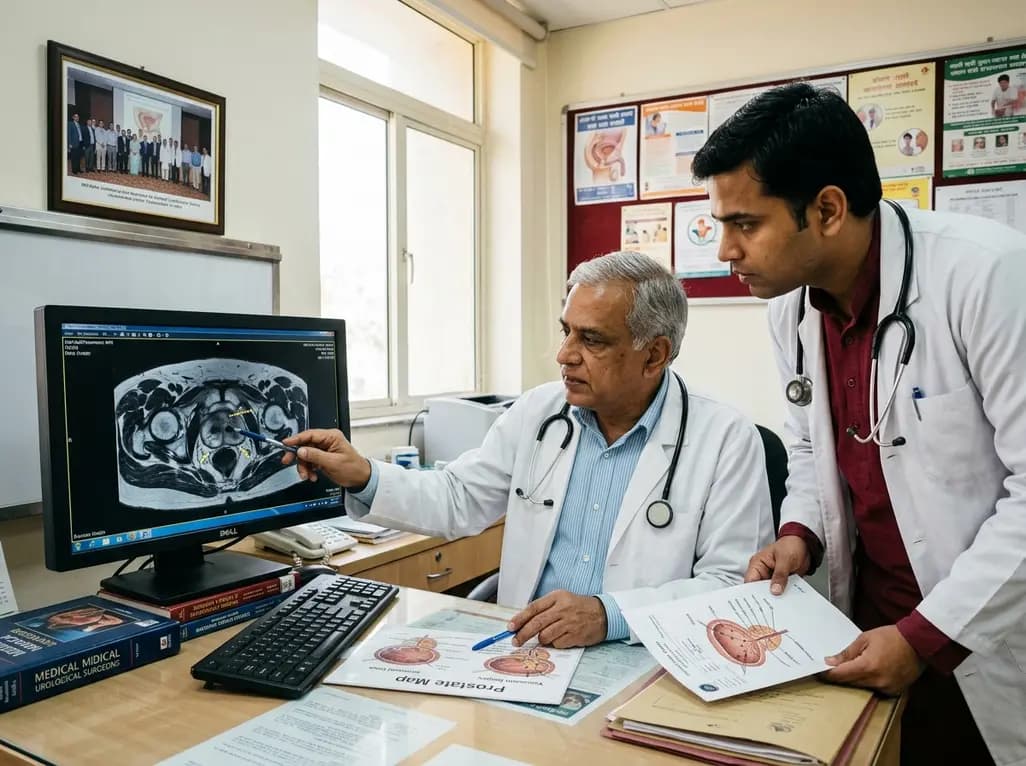
Multiparametric magnetic resonance imaging has revolutionized prostate cancer diagnostics across modern urology. Clinicians routinely debate whether omitting contralateral systematic biopsy compromises staging accuracy when performing targeted sampling for unilateral index lesions. Traditionally, international protocols have advocated combining targeted biopsy with standard twelve-core systematic sampling across both lobes. However, harvesting extensive bilateral cores increases operative time, patient discomfort, and biopsy-related complications such as infection or bleeding. Therefore, urologists increasingly seek strategies that streamline diagnostic pathways without missing high-grade malignancy. Recent clinical investigations have specifically evaluated whether targeted biopsy paired solely with ipsilateral systematic biopsy provides sufficient concordance with whole-mount radical prostatectomy pathology. Understanding the exact risk of clinically significant histological upgrading remains essential for surgical planning, active surveillance selection, and nerve-sparing decisions.
In contemporary urological practice, MRI-transrectal ultrasound fusion biopsy enhances the detection of clinically significant prostate cancer while minimizing the diagnosis of indolent disease. When an MRI demonstrates a discrete unilateral suspicious lesion, sampling the ipsilateral prostate tissue directly addresses the primary tumor volume. Nevertheless, omitting contralateral systematic biopsy cores raises legitimate concerns regarding occult, multifocal lesions residing in the radiologically negative lobe. Retrospective paired-design studies comparing full bilateral sampling against ipsilateral-only systematic approaches provide critical clarity on this dilemma. Investigators reconstruct different diagnostic pathways within identical patient cohorts undergoing definitive radical prostatectomy. Consequently, researchers can accurately assess whole-mount pathological specimens against preoperative histological grading. These analyses consistently demonstrate that ipsilateral systematic sampling captures the vast majority of clinically significant tumors. Furthermore, sparing the contralateral lobe substantially decreases core numbers without introducing unacceptable diagnostic blind spots for practicing surgeons.
Pathological concordance between preoperative biopsy and definitive surgical specimens represents the gold standard for assessing diagnostic fidelity. A primary concern when reducing biopsy templates is clinically significant upgrade, where low-grade disease (Grade Group 1) shifts to intermediate or high-grade malignancy (Grade Group 2 or higher) at radical prostatectomy. Evidence reveals that adding targeted biopsy to ipsilateral systematic cores yields pathological concordance rates comparable to full bilateral systematic sampling. Although bilateral sampling detects occasional low-volume contralateral foci, these secondary lesions rarely alter the dominant Grade Group of the prostatectomy specimen. Moreover, isolated contralateral systematic cores primarily detect indolent Grade Group 1 tumors that do not impact overall oncological risk stratification. Thus, the absolute rate of clinically significant upgrading remains stable whether urologists sample the contralateral lobe or omit it entirely in patients with unilateral index lesions.
Accurate preoperative grading directly governs radical prostatectomy planning, especially regarding nerve-sparing surgery and extended pelvic lymph node dissection. When urologists evaluate patients with unilateral disease on multiparametric MRI, knowing the true pathological status of the contralateral neurovascular bundle is crucial. Omitting contralateral biopsy cores rarely alters the contralateral nerve-sparing strategy because surgical decisions depend predominantly on MRI tumor location, capsular contact length, and ipsilateral core pathology. Additionally, nomograms estimating the risk of lymph node metastasis rely heavily on the highest biopsy Grade Group and serum prostate-specific antigen levels rather than total positive core counts in benign-appearing contralateral zones. Consequently, a refined biopsy template consisting of targeted biopsy plus ipsilateral systematic sampling preserves therapeutic precision while streamlining patient evaluation prior to robotic or open radical prostatectomy.
In Indian healthcare settings, optimizing prostate biopsy protocols offers meaningful clinical and socioeconomic advantages. Many tertiary centers across India now utilize high-resolution multiparametric MRI and cognitive or software-based fusion biopsy platforms. However, healthcare resources vary widely between urban centers and regional facilities. Reducing unnecessary biopsy cores directly lowers the procedural burden, shortens anesthesia exposure, and decreases consumable costs for pathology processing. Furthermore, transperineal biopsy approaches are rapidly gaining popularity in India due to near-zero sepsis rates compared to transrectal sampling. Applying a targeted plus ipsilateral template aligns perfectly with transperineal workflows by minimizing puncture trauma and postoperative urinary retention. Nevertheless, clinicians must ensure high-quality imaging interpretation and rigorous radiological-pathological correlation before selectively de-escalating contralateral sampling in Indian patients presenting with elevated prostate-specific antigen levels.
Every prostate biopsy template involves an inherent trade-off between maximizing diagnostic yields and minimizing procedural morbidity. Extended saturation biopsies undoubtedly sample more tissue, but they simultaneously escalate patient discomfort, transient hematuria, acute urinary retention, and infectious complications. By safely omitting contralateral systematic cores in well-characterized unilateral index lesions, urologists achieve an optimal middle ground. This focused approach maintains high diagnostic sensitivity for clinically meaningful malignancies while shielding patients from the physical harms of unnecessary tissue trauma. As imaging resolution and artificial intelligence-assisted lesion detection continue to advance, diagnostic templates will become increasingly tailored to individual risk profiles. Modern evidence strongly supports this patient-centric de-escalation, reassuring clinicians that omitting non-targeted contralateral cores does not compromise oncological safety or surgical outcomes.
Clinical evidence indicates that omitting contralateral systematic biopsy in patients with unilateral MRI-visible lesions does not significantly increase clinically meaningful upgrading at radical prostatectomy. While bilateral sampling occasionally detects additional microscopic foci, these secondary lesions are predominantly low-grade and rarely alter the overall pathological Grade Group or definitive oncological staging.
Combining MRI-targeted biopsy with ipsilateral systematic biopsy reduces total tissue core harvesting, shortens procedural time, and lowers post-biopsy complications such as hematuria, acute urinary retention, and infection. Furthermore, this streamlined approach preserves diagnostic accuracy for clinically significant prostate cancer without subjecting patients to unnecessary bilateral sampling trauma.
Surgical planning for nerve-sparing prostatectomy relies primarily on multiparametric MRI findings, dominant index lesion localization, and ipsilateral biopsy pathology. Omitting contralateral systematic biopsies rarely alters contralateral nerve-sparing decisions or indications for pelvic lymph node dissection, thereby maintaining excellent functional and oncological outcomes for surgical candidates.
Disclaimer: This content is for informational and educational purposes only and does not constitute medical advice. Consult a qualified healthcare provider for diagnosis and treatment. Refer to the latest local and national guidelines for clinical practice.
References
Jin S et al. Impact of omitting contralateral systematic biopsy on clinically significant upgrading at radical prostatectomy: a retrospective paired-design study. Int Urol Nephrol. 2026 Aug 23. doi: 10.1007/s11255-026-05344-5. PMID: 42633607.
Ahdoot M, Wilbur AR, Reese SE, et al. MRI-Targeted, Systematic, and Combined Biopsy for Prostate Cancer Diagnosis. N Engl J Med. 2020;382(10):917-928.
Ploussard G, Roumiguié M, Rozet F, et al. The Impact of Omitting Contralateral Systematic Biopsy on the Surgical Planning of Patients with a Unilateral Suspicious Lesion on Magnetic Resonance Imaging Undergoing Robot-assisted Radical Prostatectomy for Prostate Cancer. Eur Urol Open Sci. 2024;62:101-108.

Read summarized clinical updates, watch expert medical content, and earn CME certifications right from your smartphone.
Open your smartphone camera to scan and install.





Investigating whether omitting contralateral systematic biopsy during MRI-targeted fusion biopsy increases clinically significant upgrading at radical prostatectomy, this study provides vital insights for optimizing prostate cancer diagnosis and surgical planning.
last month

Chronic eosinophilic pneumonia can present with atypical bronchoalveolar lavage findings and an organizing pneumonia pattern, necessitating surgical lung biopsy.
Today

Discover the crucial distinctions between mesonephric and mesonephric-like proliferations of the female genital tract. Learn the key histomorphologic, immunohistochemical, and molecular differences essential for accurate diagnosis and clinical management.
Today
Kikuchi-Fujimoto disease rarely presents with refractory shock and sinus bradycardia in children. This case report highlights life-threatening hemodynamic instability requiring intensive care, preserved cardiac contractility, and the critical diagnostic role of early excisional lymph node biopsy.
Today

A narrative review investigates whether the articularis genus muscle functions as an independent anatomical entity or blends with the vastus intermedius. We evaluate its morphology, role in retracting the suprapatellar bursa, and direct clinical significance in anterior knee pain and arthroplasty.
Today

A Bayesian multilevel meta-analysis reveals that aerobic training combined with moderate carbohydrate restriction modestly lowers HbA1c in type 2 diabetes. However, sparse data and very low certainty leave incremental benefits over exercise or diet alone unproven, highlighting the need for individualized care.
Today
