.webp&w=384&q=75)

Loading, please wait...
Loading, please wait...

Glioblastoma represents the most aggressive primary brain malignancy encountered in neuro-oncology practice today. Historically, clinicians identified these lethal neoplasms by intense ring enhancement and central necrosis on magnetic resonance imaging. However, modern clinical practice increasingly uncovers atypical radiological presentations that challenge classic diagnostic concepts. Specifically, non-contrast-enhancing glioblastoma exhibits a deceptive, low-grade radiologic appearance while maintaining an aggressive IDH-wildtype molecular profile. Consequently, multidisciplinary teams must recognize how these unique tumors deviate from conventional diagnostic patterns. Furthermore, prompt recognition enables clinicians to initiate aggressive, life-extending therapeutic strategies without delay.
Clinicians typically associate IDH-wildtype glioblastomas with thick ring enhancement and robust microvascular breakdown. In contrast, non-contrast-enhancing glioblastoma defies this canonical imaging appearance. These atypical neoplasms present as diffuse T2 and fluid-attenuated inversion recovery signal hyperintensities lacking significant gadolinium uptake. Therefore, radiologists and clinicians often mistake them for lower-grade diffuse astrocytomas during initial evaluations.
A seminal study from Heidelberg University Hospital systematically analyzed 352 patients with confirmed IDH-wildtype glioblastoma. Using a volumetric threshold of one cubic centimeter, investigators classified 12.5% of tumors as non-enhancing. Consequently, this study demonstrated that an intact blood-brain barrier does not preclude highly malignant biology. In addition, patients in this cohort presented at a slightly younger age with seizures rather than focal neurologic deficits. Because these tumors mimic benign lesions, healthcare providers must maintain heightened clinical vigilance. Thus, diagnostic teams can avoid dangerous observational delays.
Pathologists traditionally evaluate surgical tissue for distinct microstructural hallmarks to establish a glioblastoma diagnosis. Specifically, prominent microvascular proliferation and pseudopalisading necrosis represent the diagnostic bedrock of grade 4 astrocytomas. However, non-enhancing tumors exhibit profound morphological divergence from these criteria. The Heidelberg researchers found microvascular proliferation in only 39% of non-enhancing specimens, compared to 94% of enhancing glioblastomas. Similarly, tissue necrosis appeared in only 25% of non-enhancing tumors versus 92% of enhancing controls.
Furthermore, cellular proliferation rates differ markedly between these radiographic phenotypes. Enhancing tumors display intense mitotic activity with a mean Ki-67 proliferation index of 26%. In contrast, non-enhancing glioblastomas demonstrate a significantly lower Ki-67 index of 18%. Because these primary microscopic hallmarks are frequently absent, routine light microscopy often causes diagnostic confusion. Consequently, pathologists can easily misinterpret these aggressive tumors as low-grade diffuse gliomas. Therefore, diagnostic laboratories cannot rely on histopathology alone to rule out glioblastoma.
Because classic histopathology frequently underestimates tumor grade, molecular testing provides indispensable diagnostic certainty. The 2021 World Health Organization classification updated glioma grading by integrating objective genetic markers. Under these guidelines, an IDH-wildtype diffuse astrocytoma receives a glioblastoma diagnosis if specific molecular alterations are present. These crucial biomarkers include TERT promoter mutations, EGFR gene amplification, and concurrent chromosome 7 gain and chromosome 10 loss.
Notably, the Heidelberg study revealed that 55% of non-enhancing glioblastoma cases received their diagnosis solely through molecular criteria. In stark contrast, only 3% of contrast-enhancing tumors required molecular profiling to confirm glioblastoma status. Therefore, molecular testing represents the single most vital diagnostic bridge for identifying atypical glioblastomas. In addition, next-generation sequencing prevents devastating diagnostic delays that could compromise therapeutic timing. Ultimately, modern neuro-oncology requires routine molecular assessment for every non-enhancing adult diffuse glioma.
The distinct biological characteristics of non-enhancing glioblastomas directly affect patient survival. Contrast-enhancing glioblastomas carry a notoriously poor prognosis, yielding a median overall survival of 14.7 months. In contrast, patients with non-enhancing glioblastomas achieved a median overall survival of 27.2 months in the Heidelberg cohort. This statistically significant survival benefit underscores the less aggressive immediate biology of these tumors.
Moreover, multivariate analyses confirmed the absence of contrast enhancement as an independent favorable prognostic marker. Lower mitotic rates and an intact blood-brain barrier likely indicate an early developmental evolutionary phase. Consequently, clinicians can approach patient discussions with realistic, measured hope. However, physicians must recognize that these tumors retain aggressive IDH-wildtype genetics. Over time, many non-enhancing lesions eventually develop robust contrast enhancement, tissue necrosis, and accelerated clinical deterioration. Therefore, oncologists must balance optimism with rigorous, ongoing surveillance.
These unique radiological and histological traits significantly reshape neurosurgical strategy and adjuvant treatment planning. For classic glioblastomas, neurosurgeons pursue maximal resection guided by gadolinium-enhancing margins and intraoperative fluorescence. However, non-enhancing tumors do not accumulate fluorophores like 5-aminolevulinic acid due to an intact vascular barrier. Therefore, surgical teams must utilize intraoperative magnetic resonance imaging, neuro-navigation, and cortical mapping to achieve safe supratotal resections within FLAIR-hyperintense zones.
Furthermore, medical oncologists must deliver proactive postoperative care. Even when biopsy specimens lack visible necrosis, clinicians should administer standard chemoradiation according to the Stupp protocol. Additionally, testing for MGMT promoter methylation status helps oncologists predict sensitivity to temozolomide chemotherapy. In clinical environments with evolving molecular infrastructure, pathology departments must prioritize molecular testing for non-enhancing diffuse gliomas. Thus, multidisciplinary teams can deliver timely, aggressive therapies that maximize long-term survival.
Advanced neuroimaging tools provide essential diagnostic clarity when standard anatomical MRI sequences show equivocal findings. In contrast to conventional scans, perfusion-weighted magnetic resonance imaging evaluates microvascular alterations by measuring regional cerebral blood volume. Similarly, proton magnetic resonance spectroscopy detects elevated choline ratios and decreased N-acetylaspartate, identifying malignant metabolic activity within non-enhancing lesions.
In addition, amino acid positron emission tomography using fluoroethyl-L-tyrosine highlights occult areas of high metabolic demand. These advanced imaging techniques guide neurosurgeons toward the most active tumor regions for targeted stereotactic biopsy. Consequently, surgical teams avoid unrepresentative biopsies that cause dangerous histological misclassifications. Moreover, incorporating artificial intelligence and radiomic profiling can predict underlying IDH mutation status preoperatively. For healthcare institutions in India, adopting standardized imaging protocols and expanding molecular diagnostic access will substantially enhance clinical care. Ultimately, comprehensive multimodal evaluation ensures accurate diagnosis and optimized outcomes for glioblastoma patients.
Clinicians identify these tumors reliably by combining advanced physiological neuroimaging with mandatory molecular testing. Although non-enhancing glioblastomas resemble low-grade gliomas on standard MRI, molecular analysis reveals IDH-wildtype status alongside TERT promoter mutations, EGFR amplification, or chromosome 7/10 alterations. This genetic profile establishes an unequivocal glioblastoma diagnosis despite non-enhancing radiology.
Patients demonstrate prolonged survival because non-enhancing glioblastomas exhibit lower Ki-67 proliferative rates and an intact blood-brain barrier. These features indicate an earlier, less aggressive phase of tumor evolution. Consequently, patients achieve a median overall survival of 27.2 months, nearly double the 14.7 months observed in conventional contrast-enhancing glioblastomas.
Neurosurgeons encounter major visualization challenges because non-enhancing tumors rarely accumulate fluorophores like 5-aminolevulinic acid due to intact vascular barriers. Therefore, surgical teams rely on intraoperative magnetic resonance imaging, advanced neuro-navigation, and awake functional brain mapping to resect FLAIR-hyperintense infiltrative tumor tissue safely while preserving crucial neurological functions.
Disclaimer: This content is for informational and educational purposes only and does not constitute medical advice, diagnosis, or treatment recommendations. Clinical decisions should always be made by qualified healthcare professionals based on individual patient assessments and validated diagnostic criteria. Refer to the latest local and national guidelines for clinical practice.
References

Read summarized clinical updates, watch expert medical content, and earn CME certifications right from your smartphone.
Open your smartphone camera to scan and install.





A landmark Heidelberg study reveals that non-contrast-enhancing glioblastoma frequently lacks traditional necrosis and microvascular proliferation, relying on molecular markers for diagnosis, and demonstrates significantly longer overall survival than contrast-enhancing counterparts.
Today
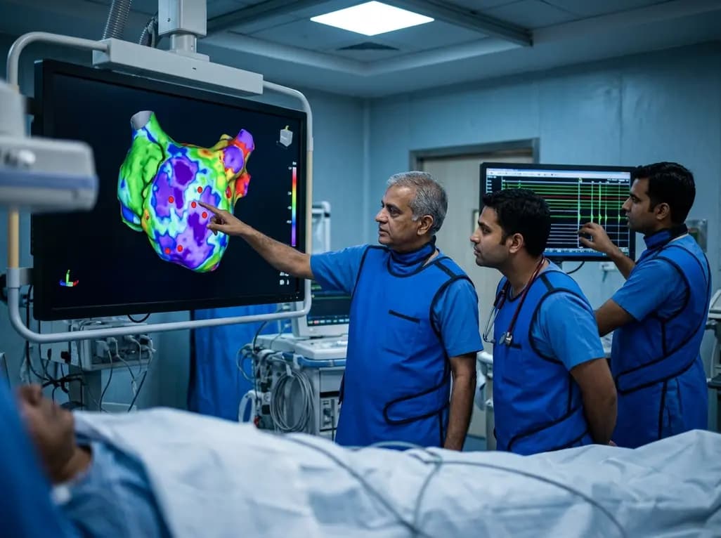
A study of 1,428 AF ablation patients reveals that over 60% of left atrial non-PV triggers arise within low-voltage areas, with anterior and roof triggers colocalizing entirely within fibrotic substrate. Identifying and ablating these sites is critical for long-term arrhythmia freedom.
Today

A comprehensive meta-analysis of over 278,000 women demonstrates that maternal atopic dermatitis significantly increases risks of premature rupture of membranes and low birth weight. Discover essential clinical insights on disease mechanisms, serial antenatal growth monitoring, and safe therapeutic interventions.
Today
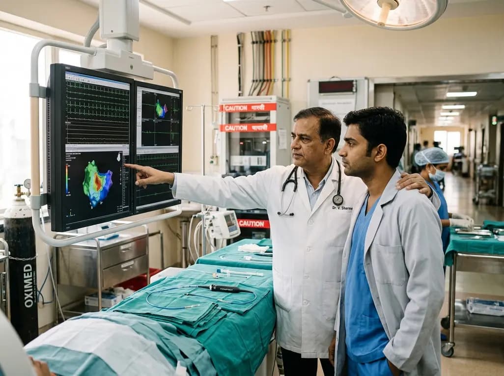
A pioneering study highlights the first United States experience using the FARAPOINT focal pulsed-field ablation catheter for refractory ventricular tachycardia. The catheter achieved complete acute non-inducibility across tested patients without procedural complications, marking a milestone in ventricular ablation.
Today

A biomechanical study shows that augmenting thin fascia lata grafts with a reinforced polyester patch doubles ultimate tensile strength and stiffness, matching the performance of thick grafts for superior capsule reconstruction while reducing donor-site morbidity.
Today

A cross-sectional study reveals that elevated METS-IR is independently associated with higher myopia odds in adolescents, showing biological synergy with obesity and highlighting the need for metabolic screening in pediatric eye care.
Today
