.webp&w=384&q=75)

Loading, please wait...
Loading, please wait...
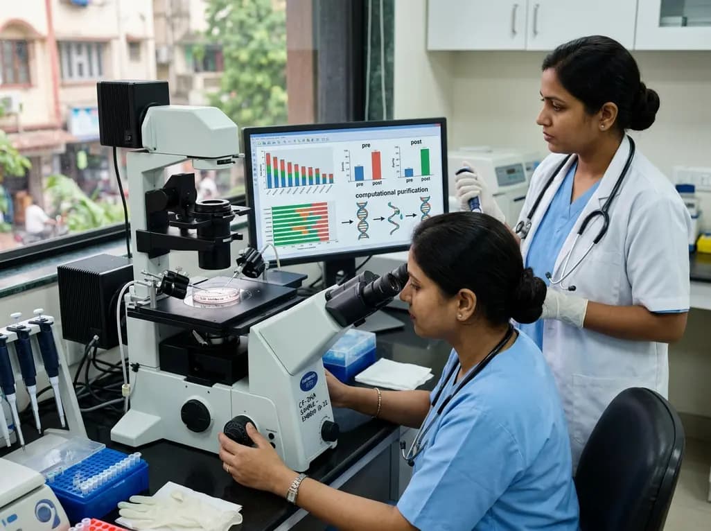
Assisted reproductive technology continues to advance toward safer, highly precise diagnostic methodologies. Clinicians have traditionally relied on morphological grading and invasive biopsies to evaluate embryo developmental competence. However, the emergence of non-invasive preimplantation genetic testing provides a transformative alternative. By analyzing spent embryo culture medium, practitioners can harvest embryo cfDNA without physically disturbing delicate blastocysts. A novel computational and diagnostic architecture, termed the NICE framework, offers significant breakthroughs in signal purification, resolving historical contamination issues and refining clinical embryo selection.
Preimplantation genetic testing for aneuploidies represents a cornerstone of modern reproductive medicine. Standard protocols require trophectoderm biopsy at the blastocyst stage, where embryologists remove several cells for molecular analysis. Although this approach yields robust genetic data, it presents notable clinical challenges. Biopsy requires advanced micromanipulation skills, expensive laser equipment, and specialized embryology training. Furthermore, physical cellular removal introduces potential procedural stress that may impair embryo implantation potential. In addition, trophectoderm biopsy may fail to represent inner cell mass composition accurately due to biological mosaicism. Consequently, reproductive specialists have actively explored non-invasive preimplantation genetic testing. By sampling spent embryo culture medium, laboratories collect cell-free nucleic acids shed naturally during embryonic cleavage. This method avoids mechanical trauma, preserves cellular architecture, and simplifies laboratory workflows. Therefore, non-invasive assessment represents an ideal screening paradigm for broader clinical adoption in assisted reproductive technology.
Despite the compelling advantages of spent medium analysis, critical technical obstacles have delayed widespread clinical implementation. The primary challenge involves maternal DNA contamination within the culture droplet. Residual cumulus cells frequently linger around the zona pellucida despite rigorous denudation procedures. Furthermore, degenerating polar bodies release maternal genomic fragments directly into the surrounding medium. Consequently, background maternal DNA competes with genuine embryonic signals during whole-genome amplification and next-generation sequencing. This phenomenon causes profound diagnostic discrepancies, including false-negative aneuploidy detections, missed paternal deletions, and discordant sex chromosome determinations. In clinical settings, such artifacts jeopardize patient trust and risk inappropriate embryo prioritization. In addition, low overall concentrations of cell-free fragments exacerbate amplification bias and technical noise. Therefore, developing robust computational workflows capable of isolating authentic embryonic signatures from maternal background noise remains an absolute clinical imperative.
To overcome these persistent contamination hurdles, researchers developed the NICE (Non-Invasive CfDNA-based Embryo assessment) framework. This platform utilizes a two-step strategy that combines bioinformatic purification with intelligent embryo scoring. Central to the initial purification step is DECENT-plus, an advanced deep copy number variation reconstruction algorithm. DECENT-plus builds upon previous deep-learning architectures to deconvolve mixed genomic signals mathematically. The algorithm effectively models the unique fragmentation patterns and copy-number ratios characteristic of polar body and cumulus contaminants. By differentiating maternal noise from true embryonic reads, DECENT-plus isolates authentic embryonic genomic data with superior resolution. Furthermore, this computational deconvolution eliminates the need for complex, destructive physical separation techniques. As a result, the framework salvages low-yield sequencing runs that conventional pipelines would otherwise discard as uninformative. This algorithmic purification establishes a reliable foundation for downstream genetic evaluation and clinical decision-making.
Following genomic purification, the NICE framework executes its second phase: machine-learning-driven quality classification. Raw sequencing metrics and biometric features extracted from the purified nucleic acids feed directly into predictive algorithms. These features include fragment-length distributions, chromosomal stability scores, and quantitative copy-number variations across the embryonic genome. Machine learning models analyze these multidimensional data points simultaneously to determine developmental viability. By moving beyond binary ploidy calls, the system generates standardized, objective embryo viability indexes. Consequently, reproductive endocrinologists and embryologists receive intelligent decision support that complements traditional time-lapse morphokinetics. This integrated assessment enables prioritized transfer of blastocysts with the highest implantation and live-birth potential. Moreover, the standardized scoring reduces inter-operator variability across different embryology laboratories, promoting uniform clinical outcomes and enhancing overall laboratory consistency.
The clinical implementation of the NICE framework offers transformative benefits for modern in vitro fertilization practice. First, non-invasive collection streamlines laboratory logistics by removing the scheduling bottlenecks associated with micro-biopsies. Laboratory technicians simply collect culture droplets during routine embryo handling or prior to cryopreservation. Second, this workflow significantly reduces consumables costs, making genetic screening accessible to a broader patient demographic. Third, avoiding biopsy eliminates freeze-all mandates driven solely by biopsy recovery times, thereby expanding fresh transfer opportunities when clinically appropriate. Additionally, the improved accuracy delivered by DECENT-plus mitigates the risk of discarding viable mosaic embryos prematurely. ART programs can integrate these bioinformatic pipelines directly into existing next-generation sequencing infrastructure without purchasing specialized hardware. Accordingly, this technological leap bridges the long-standing gap between experimental non-invasive testing and routine, high-throughput clinical practice.
As non-invasive embryo testing matures, reproductive medicine must address vital ethical, regulatory, and technical considerations. Clinicians must provide comprehensive pre-test counseling regarding the screening nature of cell-free DNA analysis. Because non-invasive testing evaluates extracellular material, patients must understand that definitive diagnostic confirmation may still require prenatal screening during gestation. Furthermore, standardized international validation trials remain essential to benchmark the NICE framework across diverse culture media formulations and incubation conditions. In the future, combining cell-free DNA profiling with spent-medium metabolomics, microRNA signatures, and artificial intelligence-driven optical imaging will create comprehensive multi-omic embryo assessments. Such multimodal platforms will dramatically enhance personalized embryo selection algorithms. Ultimately, frameworks like NICE represent a decisive step toward fully automated, safe, and highly reliable non-invasive embryology workflows that maximize pregnancy rates worldwide.
Embryonic cell-free DNA enters the spent culture medium through natural cellular processes, including physiological apoptosis, shedding of cellular fragments, and blastocoele fluid leakage during blastocyst expansion. As embryonic blastomeres divide and undergo cellular turnover during preimplantation development, genomic fragments leach into the extracellular environment. Consequently, embryologists can sample this fluid non-invasively to assess genetic integrity without performing physical micro-biopsies on delicate embryonic blastomeres.
Maternal contamination predominantly originates from residual maternal cumulus cells or persistent polar body fragments attached to the zona pellucida. These maternal genomic materials release competing cell-free DNA into the spent medium during culture. Consequently, standard sequencing pipelines may amplify maternal reads instead of embryonic signals. This contamination often produces false-negative results, obscures paternal chromosomal contributions, and causes discordant sex determination during preimplantation genetic screening.
The NICE framework utilizes the DECENT-plus algorithm to computationally eliminate maternal background signals and polar body artifacts from raw sequencing data. Next, it extracts multi-dimensional biometric features from the purified embryonic DNA and processes them through machine learning classifiers. As a result, reproductive teams obtain objective, standardized viability predictions and aneuploidy risk assessments, enabling safe and non-invasive embryo prioritization before clinical transfer.
Disclaimer: This content is for informational and educational purposes only and does not constitute medical advice or establish a doctor-patient relationship. Healthcare professionals must exercise their independent clinical judgment. While based on verified clinical sources, the author and publisher disclaim liability for any errors, omissions, or clinical decisions made based on this material. Refer to the latest local and national guidelines for clinical practice.
References
1. Zhou X et al. NICE: A Two-Step Non-Invasive Framework for Embryo cfDNA Read Enrichment and Quality Assessment. Adv Sci (Weinh). 2026 Aug 24. doi: 10.1002/advs.77327. PMID: 42635625.
2. Rubio C, et al. Embryonic cell-free DNA in spent culture medium: a non-invasive tool for aneuploidy screening. Fertil Steril. 2019;112(3):e26.
3. Huang L, et al. Non-invasive preimplantation genetic testing of embryonic genome in spent culture medium. Hum Reprod. 2023;38(6):1120-1132.

Read summarized clinical updates, watch expert medical content, and earn CME certifications right from your smartphone.
Open your smartphone camera to scan and install.





The NICE framework introduces a two-step non-invasive approach combining DECENT-plus computational purification and machine learning to analyze embryo cfDNA from spent culture medium, overcoming maternal contamination and enabling accurate embryo prioritization in assisted reproductive technology.
last month

A nationwide English cohort study shows that interstitial lung diseases are no longer rare, with incidence jumping 30% and 200,000 active cases. This analysis highlights clinical implications for diagnosis, multi-disciplinary management, and global health priorities across respiratory and internal medicine.
Today

A nationwide South Korean study shows underreporting of smoking among women dropped from 57.4% to 36.4% between 2008 and 2021. However, over one-third of female smokers still conceal their habit, highlighting the critical role of cotinine verification in clinical risk assessment.
Today

PepsiCo and Monster Beverage have challenged the FSSAI ban on the energy drink label in Indian courts. While companies highlight supply disruptions, regulatory actions spotlight severe cardiovascular and metabolic risks associated with excessive caffeine, sugar, and taurine consumption, especially in adolescents.
Today

A new study evaluates the rheological stability of decellularized peripheral nerve matrix hydrogels under varied storage conditions. Findings show -80°C storage and freeze-drying preserve gelation kinetics and mechanical stiffness, offering key insights for shipping and translational clinical applications.
Today

A Bayesian multilevel meta-analysis reveals that aerobic training combined with moderate carbohydrate restriction modestly lowers HbA1c in type 2 diabetes. However, sparse data and very low certainty leave incremental benefits over exercise or diet alone unproven, highlighting the need for individualized care.
Yesterday
