.webp&w=384&q=75)

Loading, please wait...
Loading, please wait...

Mechanical thrombectomy has revolutionized acute ischemic stroke management across emergency centers globally. However, evaluating post-thrombectomy outcomes remains a formidable clinical challenge because angiographic recanalization does not always guarantee neurological recovery. Although clinicians frequently achieve modified Treatment in Cerebral Ischemia scores of 2b or higher, reperfusion injury and microvascular compromise frequently hamper functional restoration. Therefore, neurointensivists require objective bedside tools to detect impending clinical deterioration during the earliest post-procedural phase. Integrating clinical risk factors with physiological biomarkers offers an exceptional pathway to identify high-risk patients who require intensified monitoring and targeted therapeutic interventions.
Endovascular thrombectomy achieves excellent macrovascular reopening in most individuals presenting with anterior circulation large vessel occlusion. Despite achieving complete or near-complete recanalization, nearly half of treated individuals fail to regain functional independence at 90 days. Clinicians refer to this frustrating clinical paradox as futile reperfusion. Several pathophysiological processes contribute to this disconnect between vessel patency and functional recovery. Specifically, microvascular no-reflow, breakdown of the blood-brain barrier, and downstream microthrombi severely restrict cellular oxygen delivery. Moreover, impaired cerebral autoregulation can trigger severe hyperperfusion syndrome or hemorrhagic transformation. Consequently, relying solely on immediate post-procedural catheter angiography provides an incomplete assessment of cerebral tissue viability. Standard clinical assessments like the National Institutes of Health Stroke Scale offer valuable tracking, yet they often lag behind silent hemodynamic compromise. In addition, delayed cerebral edema may advance rapidly before overt clinical signs manifest at the bedside. Therefore, bedside monitoring tools that capture dynamic intracranial blood flow changes are vital for neurocritical care teams. Developing precise risk models can effectively identify vulnerable patients who require tailored blood pressure targets, continuous neuromonitoring, and neuroprotective strategies during neurointensive care.
To bridge this diagnostic gap, researchers developed an innovative multimodal prognostic model evaluating 90-day post-thrombectomy outcomes. This clinical cohort study examined 176 patients presenting with anterior circulation large vessel occlusion who underwent successful mechanical thrombectomy. Each patient underwent systematic transcranial Doppler ultrasonography within 72 hours following the endovascular procedure. Investigators randomly assigned participants into training and validation cohorts using a standard seven-to-three allocation ratio. Subsequently, the research team applied least absolute shrinkage and selection operator regression analysis to eliminate collinearity and identify dominant prognostic indicators. The analysis identified three critical determinants: peak systolic velocity ratio, mean flow velocity of the affected middle cerebral artery, and active smoking status. Furthermore, receiver operating characteristic curve analysis confirmed that the integrated multimodal nomogram achieved exceptional discrimination. Calibration plots demonstrated high agreement between predicted functional trajectories and observed 90-day modified Rankin Scale scores. Additionally, decision curve analysis verified meaningful clinical net benefit across practical decision thresholds, confirming the model's bedside reliability. These findings underscore the clinical value of combining dynamic physiological measurements with readily accessible patient characteristics.
Transcranial Doppler ultrasonography provides real-time, non-invasive assessment of intracranial hemodynamics directly at the bedside. In this predictive model, both the peak systolic velocity ratio and the mean flow velocity of the affected middle cerebral artery emerged as pivotal hemodynamic markers. Specifically, an elevated peak systolic velocity ratio indicates asymmetric flow resistance and localized focal stenosis within the recanalized intracranial territory. When post-intervention arteries experience persistent mural irregularity or residual platelet aggregation, systolic blood velocity increases markedly. Conversely, an abnormally suppressed mean flow velocity within the affected middle cerebral artery indicates downstream microcirculatory collapse or high downstream vascular resistance. In addition, profound microvascular spasm can restrict forward diastolic flow, reducing overall mean perfusion velocity. Thus, sonographic evaluation captures ongoing microvascular failure that conventional angiographic imaging readily misses. By obtaining bedside hemodynamic measures within the crucial 72-hour postoperative window, intensivists can recognize impending hyperperfusion injury or early reocclusion before irreversible neurological injury develops. These measurements also provide clinicians with an objective basis for adjusting vasopressor support and individualized mean arterial pressure goals.
Beyond sonographic hemodynamic markers, cigarette smoking was identified as an independent clinical risk factor within the final multivariable model. Although some historical literature discussed a controversial neurovascular smoking paradox, modern clinical data consistently link chronic tobacco exposure to poorer functional recovery. Specifically, chronic cigarette smoke exposure induces profound systemic and cerebrovascular endothelial dysfunction. Cigarette toxins trigger sustained oxidative stress, upregulate circulating inflammatory cytokines, and impair nitric oxide bioavailability. Consequently, cerebral autoregulation suffers severe disruption, rendering the newly reperfused brain tissue highly vulnerable to fluctuating systemic blood pressure. Furthermore, smoking promotes platelet hyperreactivity, alters blood viscosity, and impairs endogenous fibrinolysis. These hematological alterations directly increase the risk of microvascular thrombosis within small downstream penetrating arterioles. Therefore, active smokers experience blunted capillary reperfusion even when macrovascular thrombectomy completely clears the primary occlusion. Identifying smoking history alongside ultrasound abnormalities allows neurocritical clinicians to anticipate heightened neuroinflammatory cascades and delayed cellular healing. Recognizing these persistent risks encourages proactive lifestyle education and aggressive secondary prevention protocols early during hospital admission.
Integrating bedside vascular ultrasound with clinical risk profiling offers significant practical advantages for hospital stroke units. In acute clinical settings, transcranial Doppler provides a cost-effective, repeatable, and non-invasive modality that avoids transporting unstable patients to radiology suites. Furthermore, combining flow velocities with patient-specific history empowers multidisciplinary teams to personalize post-recanalization hemodynamic goals. For instance, individuals identified as high-risk through elevated velocity ratios or suppressed flow parameters may benefit from stricter blood pressure control to prevent catastrophic secondary reperfusion hemorrhage. Similarly, discovering persistent distal flow stagnation may prompt heightened antiplatelet surveillance, close neuromonitoring, or specialized neuroprotective interventions. Nevertheless, clinicians must interpret these ultrasound parameters within the broader clinical context, acknowledging potential operator variability and anatomical acoustic window limitations. In resource-limited hospital settings, implementing portable vascular Doppler units can democratize post-thrombectomy surveillance where serial computed tomography is unavailable. As neurointerventional techniques continue to evolve rapidly, implementing validated predictive models will ensure that technical angiographic success translates reliably into tangible, long-term functional recovery for acute stroke survivors.
Transcranial Doppler provides continuous, bedside hemodynamic evaluation following mechanical thrombectomy. It non-invasively tracks intracranial blood flow velocities, enabling clinicians to identify downstream vascular resistance, microvascular spasm, persistent residual stenosis, and cerebral hyperperfusion early. Consequently, it delivers functional hemodynamic insights that standard angiographic imaging cannot capture.
Smoking induces chronic endothelial dysfunction, amplifies neuroinflammation, and impairs cerebral autoregulation. Furthermore, tobacco toxins promote platelet hyperreactivity and microvascular thrombosis in downstream penetrating arterioles. These systemic and vascular disruptions collectively impair microcirculatory perfusion, which substantially undermines tissue healing despite successful mechanical reopening of large occluded arteries.
The peak systolic velocity ratio compares blood flow speed across affected and unaffected vessels. An elevated ratio signals focal vascular narrowing, dynamic mural irregularities, or asymmetric intracranial vascular resistance. Identifying these velocity discrepancies within 72 hours helps neurocritical care teams detect high-risk individuals requiring intensive hemodynamic management and surveillance.
Disclaimer: This content is for informational and educational purposes only... Refer to the latest local and national guidelines for clinical practice.
References

Read summarized clinical updates, watch expert medical content, and earn CME certifications right from your smartphone.
Open your smartphone camera to scan and install.





A newly developed prognostic model combines transcranial Doppler ultrasound metrics and smoking history to predict 90-day functional recovery in acute ischemic stroke patients following successful mechanical thrombectomy.
Today
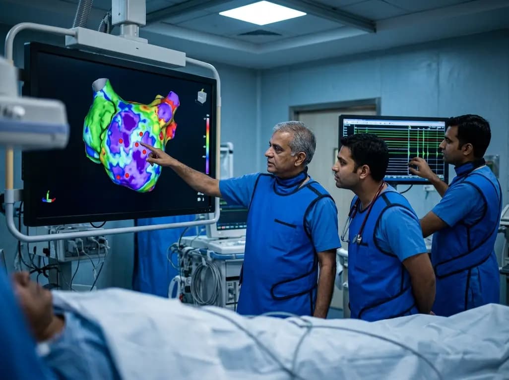
A study of 1,428 AF ablation patients reveals that over 60% of left atrial non-PV triggers arise within low-voltage areas, with anterior and roof triggers colocalizing entirely within fibrotic substrate. Identifying and ablating these sites is critical for long-term arrhythmia freedom.
Today

A comprehensive meta-analysis of over 278,000 women demonstrates that maternal atopic dermatitis significantly increases risks of premature rupture of membranes and low birth weight. Discover essential clinical insights on disease mechanisms, serial antenatal growth monitoring, and safe therapeutic interventions.
Today

A pioneering study highlights the first United States experience using the FARAPOINT focal pulsed-field ablation catheter for refractory ventricular tachycardia. The catheter achieved complete acute non-inducibility across tested patients without procedural complications, marking a milestone in ventricular ablation.
Today

A biomechanical study shows that augmenting thin fascia lata grafts with a reinforced polyester patch doubles ultimate tensile strength and stiffness, matching the performance of thick grafts for superior capsule reconstruction while reducing donor-site morbidity.
Today

A cross-sectional study reveals that elevated METS-IR is independently associated with higher myopia odds in adolescents, showing biological synergy with obesity and highlighting the need for metabolic screening in pediatric eye care.
Today
