

Loading, please wait...
Loading, please wait...

Managing acute basilar artery occlusion represents one of the most critical emergencies in modern neurovascular medicine. Posterior circulation ischemic strokes often present with fluctuating, atypical symptoms that complicate early triage. Consequently, clinicians must act swiftly to prevent catastrophic brainstem infarction and permanent disability. For many years, specialists debated whether magnetic resonance imaging or computed tomography should serve as the preferred initial diagnostic modality. Magnetic resonance imaging provides superior soft-tissue resolution for posterior fossa structures. Furthermore, diffusion-weighted imaging detects early ischemia more accurately than noncontrast computed tomography. However, magnetic resonance imaging frequently introduces logistical bottlenecks during hyperacute presentations. Patients with severe neurological deficits often require emergency intubation or metal screening before entering the scanner. Therefore, many emergency teams rely primarily on computed tomography and computed tomography angiography to accelerate treatment decisions. While computed tomography eliminates lengthy pre-scan screening, physicians have worried that it might misjudge tissue viability. Understanding whether advanced baseline imaging truly alters long-term clinical outcomes remains essential for refining global stroke protocols.
To resolve this enduring debate, investigators conducted a post hoc analysis of the randomized ATTENTION trial. This landmark study evaluated endovascular thrombectomy versus best medical management for acute posterior circulation stroke. In this secondary investigation, researchers categorized 340 enrolled patients into two cohorts according to their initial neuroimaging evaluation. Specifically, 277 patients underwent computed tomography, whereas 63 patients underwent baseline magnetic resonance imaging. The research team evaluated whether the choice between these imaging tools influenced clinical efficacy or patient safety. Importantly, both diagnostic groups presented with comparable baseline neurological severity and vascular risk profiles. Clinicians utilized noncontrast scans alongside vascular imaging to detect the arterial occlusion site and estimate tissue salvageability. Moreover, the investigators tracked procedural times, recanalization metrics, and functional recovery across both study arms. By comparing these cohorts, the authors addressed whether emergency magnetic resonance imaging provides a measurable therapeutic advantage. Consequently, the study yields robust insights into real-world neurointerventional decision-making during acute posterior circulation emergencies.
The trial primary outcome measured favorable functional recovery, defined as a modified Rankin Scale score of 0 to 3 at 90 days. Interestingly, baseline imaging modality showed no significant association with functional recovery in the overall study population. Patients evaluated with baseline magnetic resonance imaging achieved comparable odds of functional independence relative to those triaged with computed tomography. The adjusted odds ratio reached 0.99, demonstrating clinical equivalence between both diagnostic pathways. Furthermore, this lack of difference persisted consistently across both the endovascular thrombectomy group and the best medical management group. Secondary clinical endpoints also revealed remarkable alignment between the two imaging strategies. Specifically, ordinal shifts in modified Rankin Scale scores showed identical distributions between modalities. In addition, early neurological deterioration occurred at similar frequencies regardless of the initial imaging choice. Crucially, the 90-day safety outcome of all-cause mortality did not differ between the two cohorts. Therefore, selecting candidates via conventional computed tomography did not compromise long-term survival or functional independence.
Speed remains the governing principle of hyperacute stroke intervention because neuronal loss accumulates rapidly with each passing minute. Historically, clinicians assumed that performing emergency magnetic resonance imaging would impose significant operational delays. However, the ATTENTION trial data challenged this common clinical assumption. The investigators found that baseline magnetic resonance imaging did not produce statistically significant workflow delays compared to computed tomography. Specifically, the mean difference from stroke onset to trial randomization was merely 5.23 minutes between modalities. Similarly, the time from symptom onset to arterial puncture showed an insignificant mean difference of 13.16 minutes. Specialized stroke centers with streamlined magnetic resonance pathways can execute advanced imaging without sacrificing valuable procedural time. Nevertheless, establishing such rapid protocols demands substantial institutional resources, dedicated personnel, and around-the-clock scanner availability. Furthermore, the association between baseline imaging modality and clinical outcomes remained consistent across different presentation windows. Consequently, while centers can utilize magnetic resonance imaging if protocols are fast, computed tomography offers equivalent efficacy without logistical risks.
These findings deliver reassuring guidance for acute neurovascular triage in everyday clinical practice. The analysis indicates that conventional computed tomography remains fully sufficient as a first-line triage modality for acute basilar artery occlusion. Clinicians do not need to delay revascularization therapies while attempting to secure high-field magnetic resonance slots. Instead, emergency teams can confidently proceed with noncontrast computed tomography and computed tomography angiography to verify vessel occlusion. Furthermore, rapid vascular imaging confirms large vessel thrombosis while assessing posterior circulation early ischemic changes. In many community and regional hospitals, access to immediate magnetic resonance scanning remains severely restricted, especially during off-hours. Therefore, forcing an advanced imaging mandate could unintentionally deny lifesaving mechanical thrombectomy to eligible individuals. By validating computed tomography as an equally reliable selection tool, this study reinforces equitable and rapid emergency stroke care. Consequently, stroke networks should prioritize rapid image acquisition and swift intervention over complex modal selection.
In developing healthcare networks and high-volume stroke centers, optimizing resource allocation directly shapes patient survival. Computed tomography scanners operate continuously across most emergency departments, offering dependable uptime and broad accessibility. In contrast, magnetic resonance imaging facilities face frequent scheduling conflicts, longer acquisition sequences, and stringent contraindication screenings. Because patients with posterior circulation strokes frequently present in comatose or agitated states, completing motion-free magnetic resonance scans proves challenging. Furthermore, urgent airway stabilization often precludes prolonged magnetic resonance imaging suites. Implementing standardized computed tomography algorithms allows emergency physicians and neurologists to make definitive recanalization decisions within minutes of hospital arrival. Clinicians can accurately identify high-risk arterial lesions and exclude extensive established core infarction without complex perfusion software. In addition, streamlining these initial protocols shortens door-to-needle and door-to-groin puncture times across referral networks. Ultimately, adopting computed tomography-first protocols standardizes clinical workflows, reduces institutional costs, and broadens thrombectomy accessibility for diverse patient populations worldwide.
Yes. Evidence from the ATTENTION trial confirms that baseline computed tomography performs comparably to magnetic resonance imaging for selecting treatment candidates. Patients evaluated with computed tomography achieved similar 90-day functional recovery and survival rates. Therefore, conventional computed tomography remains an effective and reliable first-line imaging modality for acute basilar artery triage.
In experienced centers, magnetic resonance imaging did not cause statistically significant workflow delays. The trial demonstrated minimal mean differences in time to randomization and arterial puncture between modalities. However, because magnetic resonance imaging requires strict safety screening, computed tomography remains the faster and more practical option in most acute emergency settings.
Emergency departments should prioritize rapid noncontrast computed tomography combined with computed tomography angiography. This combination quickly excludes intracranial hemorrhage, accurately visualizes the basilar artery thrombus, and assesses posterior circulation ischemic changes. Consequently, clinicians can initiate intravenous thrombolysis or mobilize neurointerventional teams without waiting for advanced magnetic resonance imaging availability.
Disclaimer: This content is for informational and educational purposes only and is not intended as a substitute for professional medical advice, diagnosis, or treatment. Refer to the latest local and national guidelines for clinical practice.
References

Read summarized clinical updates, watch expert medical content, and earn CME certifications right from your smartphone.
Open your smartphone camera to scan and install.





A post hoc analysis of the ATTENTION trial shows that baseline CT is comparable to MRI for patient selection in acute basilar artery occlusion. Both modalities yielded similar 90-day functional recovery and safety outcomes without significant procedural delays, validating CT as an effective first-line imaging tool.
Today

The Food Safety and Standards Authority of India has issued an urgent advisory against food business operators violating mandatory labelling standards for nutraceuticals and botanicals. Regulators prohibited therapeutic claims while mandating clear advisory warnings to prevent patient toxicity and medical misuse.
Today

Biotechnology researchers in India are developing an automated liquid biopsy platform to detect cervical malignancy from blood samples. Supported by the MAHA MedTech Mission, this initiative applies nanopore sequencing and piRNA biomarker detection to eliminate invasive speculum exams and expand rural screening.
Today
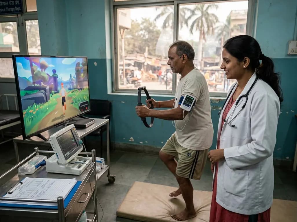
A clinical study evaluated Ring Fit Adventure in adults with chronic conditions, revealing that exergaming achieves moderate-to-vigorous exercise intensity while maintaining hemodynamic stability, high enjoyment, and zero adverse events.
Today
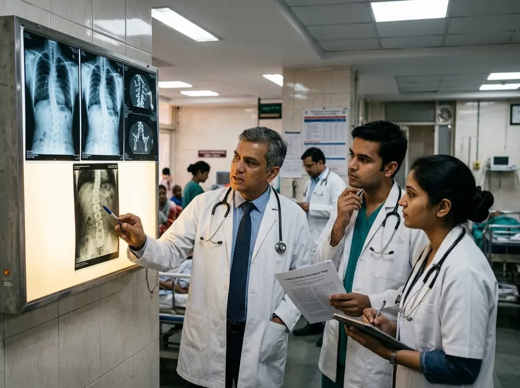
A retrospective cohort study reveals that selecting the touched vertebra as the lowest instrumented vertebra in thoracic adolescent idiopathic scoliosis yields a 20.8% distal add-on rate, driven by remnant motion segments and axial rotational discrepancies.
Today
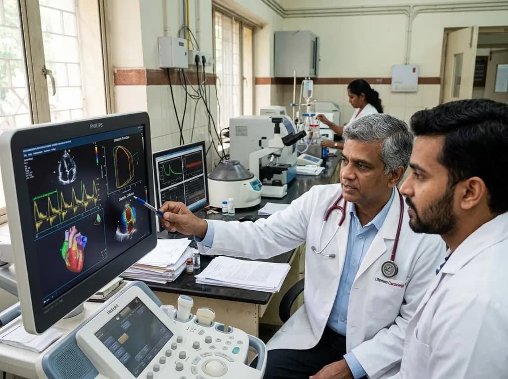
A study evaluates a cardiometabolic HFpEF model combining metabolic stress with chronic mineralocorticoid excess. The findings reveal profound diastolic stiffness, endothelial dysfunction, and pulmonary hypertension, illuminating key pathophysiological mechanisms for targeted cardiopulmonary therapies.
Today
