.webp&w=384&q=75)

Loading, please wait...
Loading, please wait...
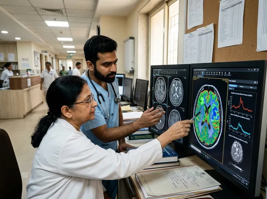
Brain metastases represent the most common intracranial malignancy in adults, frequently requiring stereotactic radiosurgery for definitive local disease control. However, distinguishing true recurrent tumor progression from treatment-induced radiation necrosis remains a formidable clinical challenge for neuro-oncologists and neuroradiologists. Because both pathological entities share overlapping radiographic features on conventional contrast-enhanced neuroimaging, clinicians frequently encounter diagnostic uncertainty. A groundbreaking study demonstrates that advanced tumor habitat analysis combining structural and physiologic magnetic resonance imaging maps provides a reliable, pathology-validated approach to resolve this critical clinical dilemma.
Stereotactic radiosurgery delivers highly focused, ablative doses of ionizing radiation to intracranial metastases while sparing adjacent normal brain parenchyma. Consequently, this intervention significantly improves local disease control and preserves cognitive function in cancer patients. Despite these therapeutic benefits, high-dose radiosurgery frequently triggers local tissue damage, producing radiation necrosis in up to one-third of treated lesions. Radiation necrosis typically manifests as an enlarging, contrast-enhancing mass surrounded by extensive vasogenic edema, closely mimicking recurrent malignant progression on standard surveillance imaging.
Standard anatomical magnetic resonance imaging cannot reliably differentiate between sterile inflammatory necrosis and aggressive neoplastic proliferation. As a result, clinicians often face profound treatment dilemmas. Misinterpreting radiation necrosis as tumor progression may expose patients to unnecessary re-irradiation or invasive surgical interventions. Conversely, mistaking viable recurrence for benign radiation necrosis delays critical systemic or targeted oncological therapies. Therefore, developing objective, non-invasive imaging biomarkers with definitive histopathological validation is imperative to guide precision neuro-oncology.
To overcome the limitations of conventional imaging, researchers developed an advanced computational framework leveraging multiparametric neuroimaging data. This methodology evaluates spatial intratumoral heterogeneity by performing unsupervised clustering across co-registered structural and functional sequences. Specifically, the processing pipeline incorporates post-contrast T1-weighted imaging, T2-weighted sequences, apparent diffusion coefficient maps from diffusion-weighted imaging, and dynamic susceptibility contrast-derived cerebral blood volume maps.
By simultaneously evaluating cellularity, membrane integrity, tissue fluid composition, and microvascular hemodynamics, the algorithm parcellates complex heterogeneous lesions into distinct subregions. Structural subregions encompass contrast-enhancing tissue, solid low-enhancing tissue, and nonviable necrotic zones. Similarly, physiologic subregions include hypervascular tissue, hypovascular cellular parenchyma, and nonviable tissue beds. Ultimately, tumor habitat analysis translates complex voxel-level multiparametric imaging signals into discrete, quantifiable sub-volumes, providing unprecedented spatial and biological insight into the microenvironment of treated brain metastases.
The investigators rigorously validated this computational technique across an independent cohort of 104 patients harboring pathologically confirmed lesions, comprising 68 recurrent metastatic tumors and 36 radiation necrosis cases. Quantitative volumetric analysis revealed striking biological differences between recurrent tumors and necrotic lesions. Recurrent metastatic lesions exhibited significantly larger contrast-enhancing volumes and higher volumes of solid low-enhancing tissue compared with radiation necrosis.
Furthermore, physiological subregions demonstrated marked differences in vascular architecture and cellular viability. Recurrent neoplasms displayed significantly higher absolute hypervascular habitat volumes and hypervascular volume fractions. Conversely, radiation necrosis lesions contained markedly higher proportions of nonviable tissue fractions across both structural and physiologic imaging paradigms. These quantitative findings directly reflect the underlying biology: recurrent metastatic tumors maintain active neoangiogenesis and dense cellular proliferation, whereas radiation necrosis primarily consists of endothelial hyalinization, fibrinoid vascular damage, and extensive coagulative tissue necrosis.
To maximize diagnostic utility in routine clinical workflows, the investigators integrated individual structural and physiologic habitat parameters into a unified composite habitat score. Receiver operating characteristic analyses demonstrated that this combined composite metric substantially outperformed individual imaging parameters alone. The combined structural and physiologic score achieved an area under the curve of 0.80 (95% confidence interval: 0.71–0.87).
Importantly, the composite habitat score yielded a high diagnostic sensitivity of 89.7% alongside a specificity of 58.3%. This robust sensitivity ensures that clinicians can reliably detect true local tumor recurrences early, minimizing the risk of untreated disease progression. In addition, the quantitative score provides an objective, reproducible numerical threshold that reduces inter-observer variability among reading radiologists. Consequently, this pathology-grounded imaging biomarker provides clinicians with actionable diagnostic confidence when evaluating ambiguous, enlarging post-radiosurgery intracranial lesions.
The successful translation of voxel-based habitat mapping into everyday neuro-oncology practice holds immense promise for optimizing individualized patient care pathways. Because this analytical technique utilizes standard multiparametric magnetic resonance sequences already acquired during routine post-treatment surveillance, clinical facilities can implement the software pipeline without requiring specialized radiotracers or additional scanner hardware. This broad accessibility represents a substantial advantage over positron emission tomography modalities in resource-conscious healthcare environments.
Moreover, integrating automated habitat segmentation into multidisciplinary tumor board discussions facilitates highly tailored therapeutic strategies. Patients identified with predominant radiation necrosis can safely avoid invasive resection and receive targeted medical management, such as corticosteroids, hyperbaric oxygen, or anti-angiogenic agents like bevacizumab. Conversely, patients identified with high-risk hypervascular recurrent tumor habitats can swiftly proceed to salvage surgical resection, laser interstitial thermal therapy, or adapted systemic therapies. As computational imaging tools continue to mature, multiparametric habitat profiling will play an indispensable role in improving survival and quality of life for patients with metastatic brain disease.
Tumor habitat analysis applies computational algorithms to multiparametric magnetic resonance images to divide heterogeneous intracranial lesions into distinct structural and physiologic subregions. This technique quantifies active cellular proliferation, microvascular perfusion, and nonviable tissue, providing objective, non-invasive biomarkers to characterize tumor biology accurately.
Both radiation necrosis and recurrent metastatic tumors disrupt the blood-brain barrier, producing identical contrast enhancement and surrounding vasogenic edema on standard anatomical magnetic resonance sequences. Conventional imaging cannot reliably separate sterile inflammatory tissue breakdown from viable malignant cellular growth without invasive histological sampling.
The composite habitat score combines structural and physiologic sub-volume metrics, achieving an area under the curve of 0.80 and 89.7% sensitivity. This high diagnostic accuracy enables multidisciplinary oncology teams to avoid unnecessary re-operations in necrosis while expediting targeted salvage therapy for recurrent disease.
Disclaimer: This content is for informational and educational purposes only. It is not intended to be a substitute for professional medical advice, diagnosis, or treatment. Always seek the advice of your physician or other qualified health provider with any questions you may have regarding a medical condition. Do not disregard professional medical advice or delay seeking it because of something you have read here. Refer to the latest local and national guidelines for clinical practice.
References
Park JE et al. Pathology-validated structural and physiologic habitat imaging for differentiating radiation necrosis from tumor recurrence in brain metastases. J Neurooncol. 2026 Aug 28. doi: 10.1007/s11060-026-05761-7. PMID: 42663779.
Smith E, Naik A, et al. Differentiating radiation necrosis from tumor recurrence: a systematic review and diagnostic meta-analysis comparing imaging modalities. J Neurooncol. 2023;162(2):291-303.
Correa R, Lei Q, et al. Advancing the characterization of enlarging lesions after stereotactic radiosurgery: The combined role of clinical timing and dosimetric profiles in distinguishing radiation necrosis from tumor recurrence. Med Phys. 2020;47(4):1725-1736.

Read summarized clinical updates, watch expert medical content, and earn CME certifications right from your smartphone.
Open your smartphone camera to scan and install.





Pathology-validated structural and physiologic MRI tumor habitat analysis provides a robust, non-invasive method to distinguish recurrent brain metastases from radiation necrosis after stereotactic radiosurgery, achieving high diagnostic sensitivity and accuracy.
4 weeks back

A nationwide English cohort study shows that interstitial lung diseases are no longer rare, with incidence jumping 30% and 200,000 active cases. This analysis highlights clinical implications for diagnosis, multi-disciplinary management, and global health priorities across respiratory and internal medicine.
Today

A nationwide South Korean study shows underreporting of smoking among women dropped from 57.4% to 36.4% between 2008 and 2021. However, over one-third of female smokers still conceal their habit, highlighting the critical role of cotinine verification in clinical risk assessment.
Today

PepsiCo and Monster Beverage have challenged the FSSAI ban on the energy drink label in Indian courts. While companies highlight supply disruptions, regulatory actions spotlight severe cardiovascular and metabolic risks associated with excessive caffeine, sugar, and taurine consumption, especially in adolescents.
Today

A new study evaluates the rheological stability of decellularized peripheral nerve matrix hydrogels under varied storage conditions. Findings show -80°C storage and freeze-drying preserve gelation kinetics and mechanical stiffness, offering key insights for shipping and translational clinical applications.
Today

A Bayesian multilevel meta-analysis reveals that aerobic training combined with moderate carbohydrate restriction modestly lowers HbA1c in type 2 diabetes. However, sparse data and very low certainty leave incremental benefits over exercise or diet alone unproven, highlighting the need for individualized care.
Yesterday
