.webp&w=384&q=75)

Loading, please wait...
Loading, please wait...

Intermediate-risk prostate cancer represents a clinically diverse group with widely variable outcomes following definitive radiation therapy. Although standard risk stratification systems classify patients into favorable and unfavorable intermediate-risk categories, biological aggressiveness often differs within these subsets. Consequently, oncologists and urologists actively seek robust imaging biomarkers to refine prognosis. Quantitative metrics such as the apparent diffusion coefficient derived from multiparametric magnetic resonance imaging offer physiological insights into tumor cellularity. Recent evidence confirms that pretreatment diffusion measurements provide meaningful prognostic information regarding long-term disease control and distant metastatic dissemination.
Multiparametric magnetic resonance imaging plays an indispensable role in contemporary prostate cancer care. Among its functional sequences, diffusion-weighted imaging evaluates the Brownian motion of water molecules within biological tissues. Densely packed malignant tissues restrict water motion significantly, which results in lower quantitative values on parametric apparent diffusion coefficient maps. Therefore, lower numerical values typically indicate higher cellular density, histological aggressiveness, and higher Gleason grades.
Historically, clinicians relied primarily on serum prostate-specific antigen values, clinical T category, and biopsy Gleason scores to categorize disease risk. However, these conventional parameters occasionally underestimate biological behavior, leading to unexpected treatment failure. Integrating quantitative diffusion measurements directly captures intratumoral biological characteristics noninvasively. Consequently, functional imaging bridges the gap between conventional anatomical evaluation and microscopic tissue behavior, providing an objective tool for pretreatment assessment.
To examine the prognostic utility of quantitative diffusion imaging, investigators analyzed a retrospective cohort of 238 men with intermediate-risk disease who underwent definitive radiotherapy. All patients completed pretreatment multiparametric magnetic resonance imaging on standardized scanners. Radiologists derived quantitative tumor measurements from baseline diffusion-weighted sequences before initiating curative-intent external beam irradiation.
Furthermore, researchers stratified the cohort according to both baseline functional imaging parameters and standard clinical risk criteria. The median follow-up extended beyond ten years, allowing adequate observation for late recurrence and metastatic spread. During this decade of surveillance, clinicians documented biochemical relapse and secondary distant progression across distinct risk groups. By tracking events over this extended timeline, the study generated robust statistical data regarding long-term cancer control. Moreover, the extended surveillance window provided meaningful clinical clarity that shorter follow-up intervals frequently fail to deliver in prostate oncology.
The long-term follow-up demonstrated twenty-two cases of biochemical failure and twelve occurrences of distant metastases across the study cohort. Notably, lower tumor apparent diffusion coefficient values, specifically defined at or below 0.668 × 10⁻³ mm²/s, strongly correlated with inferior oncologic control. Patients exhibiting these lower baseline readings experienced a significantly higher risk of developing distant metastases over time.
Statistical modeling revealed a hazard ratio of 3.94 for freedom from distant metastasis among individuals with low functional imaging metrics. Therefore, restricted water diffusion within the primary prostatic lesion served as an independent predictor of systemic progression. While local biochemical failure showed consistent patterns, distant metastatic spread demonstrated the most dramatic divergence between high and low imaging cohorts. These findings underscore that profound cellular packing within the primary nodule frequently mirrors metastatic potential, underscoring the clinical value of baseline diffusion evaluation.
When clinicians combined diffusion measurements with conventional risk stratifications, distinct prognostic subgroups emerged. Specifically, dividing patients across clinical categories and functional imaging parameters created four distinct survival tiers for both freedom from distant metastasis and freedom from biochemical failure. Patients categorized into favorable intermediate-risk with high functional diffusion values achieved excellent disease control throughout the decade.
Conversely, unfavorable intermediate-risk patients with markedly restricted tumor diffusion experienced substantially inferior outcomes. However, investigators noted that certain subsets, particularly the favorable intermediate-risk group with low diffusion readings, contained a very small sample size. Because this specific cohort included only seven patients, clinicians must interpret these granular subgroup findings as exploratory. Nevertheless, the dual-parameter approach demonstrates how quantitative functional imaging adds value to traditional risk stratification models, guiding more nuanced prognostic assessments.
Prostate cancer incidence continues to increase across India due to shifting demographic patterns, widespread diagnostic testing, and improved life expectancy. Indian tertiary cancer centers and diagnostic facilities routinely deploy advanced 3-Tesla magnetic resonance imaging platforms. Therefore, incorporating routine apparent diffusion coefficient quantification into diagnostic workflows represents an accessible, cost-effective enhancement without requiring additional invasive procedures.
Currently, treating oncologists often struggle when choosing between radiation alone and combined radiation with androgen deprivation therapy for intermediate-risk patients. Identifying tumors with pronounced diffusion restriction can help clinicians identify aggressive biology early, potentially prompting intensified therapy. However, because investigators derived the current threshold retrospectively, multicentric prospective validation remains essential before changing standard treatment protocols. Indian academic centers can lead future validation studies, refining standardized imaging protocols to optimize local radiotherapy practices.
The apparent diffusion coefficient quantifies water molecule mobility within tissue microenvironments. Lower values reflect restricted diffusion caused by high tumor cell density, nuclear atypia, and architectural distortion. Consequently, lower measurements consistently correlate with higher histological Gleason grades, increased biological aggressiveness, and elevated risks of distant metastatic dissemination following definitive radiation therapy.
Marked diffusion restriction within primary prostatic tumors correlates with significantly worse freedom from distant metastasis and biochemical control. When patients exhibit baseline values at or below established thresholds, their risk of distant relapse increases substantially. This observation suggests that restricted diffusion identifies radioresistant or biologically aggressive phenotypes requiring closer surveillance or intensified treatment strategies.
Diffusion measurements complement rather than replace established clinical classification systems such as D'Amico or National Comprehensive Cancer Network criteria. Functional imaging adds objective biological data to anatomical staging and biopsy findings. However, because current cutoffs derive from retrospective cohorts, clinicians should use diffusion data alongside conventional risk stratification tools rather than as standalone determinants.
Disclaimer: This content is for informational and educational purposes only. It does not constitute medical advice and should not substitute professional judgment. Healthcare providers must evaluate individual patient clinical contexts. Refer to the latest local and national guidelines for clinical practice.
References
Onal C et al. Prognostic Value of MRI-Derived Apparent Diffusion Coefficient in Intermediate-Risk Prostate Cancer Treated With Definitive Radiotherapy. Prostate. 2026 Sep 13. doi: 10.1002/pros.70249. PMID: 42732567.
Scialpi M, Martorana E, Scialpi P, et al. MRI apparent diffusion coefficient (ADC): A biomarker for prostate cancer after radiation therapy. Turk J Urol. 2021; 47(6): 448-451.
Pang Y, Turkbey B, Bernardo M, et al. Is apparent diffusion coefficient associated with clinical risk scores for prostate cancers that are visible on 3-T MR images? Radiology. 2011; 259(3): 775-783.

Read summarized clinical updates, watch expert medical content, and earn CME certifications right from your smartphone.
Open your smartphone camera to scan and install.





Pretreatment tumor apparent diffusion coefficient (ADC) from diffusion-weighted MRI offers vital prognostic insights in intermediate-risk prostate cancer, helping predict distant metastasis risk beyond standard clinical risk groups.
Today
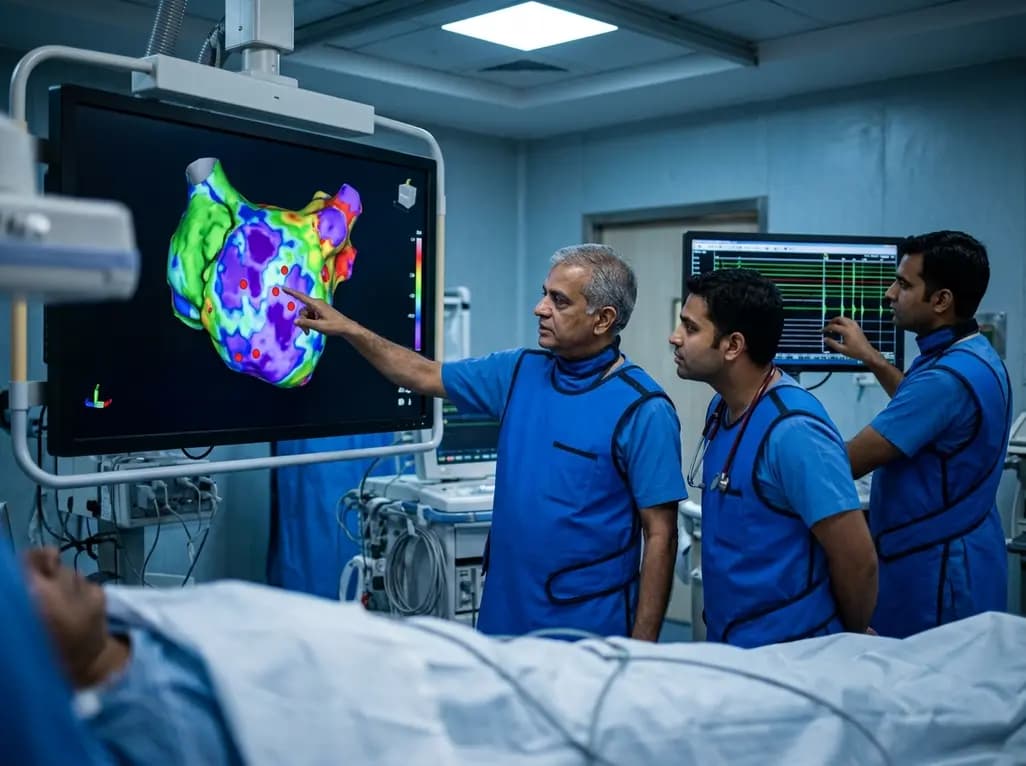
A study of 1,428 AF ablation patients reveals that over 60% of left atrial non-PV triggers arise within low-voltage areas, with anterior and roof triggers colocalizing entirely within fibrotic substrate. Identifying and ablating these sites is critical for long-term arrhythmia freedom.
Today

A comprehensive meta-analysis of over 278,000 women demonstrates that maternal atopic dermatitis significantly increases risks of premature rupture of membranes and low birth weight. Discover essential clinical insights on disease mechanisms, serial antenatal growth monitoring, and safe therapeutic interventions.
Today

A pioneering study highlights the first United States experience using the FARAPOINT focal pulsed-field ablation catheter for refractory ventricular tachycardia. The catheter achieved complete acute non-inducibility across tested patients without procedural complications, marking a milestone in ventricular ablation.
Today

A biomechanical study shows that augmenting thin fascia lata grafts with a reinforced polyester patch doubles ultimate tensile strength and stiffness, matching the performance of thick grafts for superior capsule reconstruction while reducing donor-site morbidity.
Today

A cross-sectional study reveals that elevated METS-IR is independently associated with higher myopia odds in adolescents, showing biological synergy with obesity and highlighting the need for metabolic screening in pediatric eye care.
Today
