.webp&w=384&q=75)

Loading, please wait...
Loading, please wait...
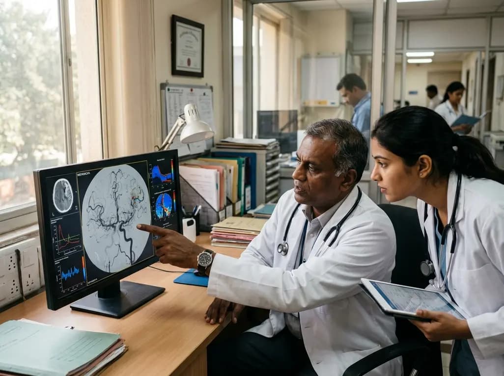
Moyamoya disease and moyamoya syndrome represent progressive intracranial steno-occlusive arteriopathies that primarily compromise the terminal internal carotid arteries and their proximal branches. Consequently, the chronic reduction in cerebral blood flow triggers compensatory collateral vascular network formation at the skull base. However, these fragile microvascular networks often fail to maintain adequate parenchymal perfusion during metabolic stress. Patients frequently present with recurrent transient ischemic attacks, disabling ischemic strokes, or catastrophic intracranial hemorrhages. Selecting the optimal strategy for moyamoya surgical revascularization remains a persistent clinical challenge because hemodynamic responses vary substantially among patients. While traditional digital subtraction angiography outlines structural vessel caliber, it fails to quantify parenchymal physiological reserve. Therefore, advanced functional neuroimaging has become indispensable for evaluating baseline cerebrovascular exhaustion. By identifying regions with severe hemodynamic compromise, clinicians can better stratify stroke vulnerability and customize procedural pathways. In particular, assessing cerebrovascular reserve helps neurosurgeons determine whether a patient requires immediate flow augmentation or indirect synangiosis. As neurovascular research advances, non-invasive physiological biomarkers increasingly guide revascularization planning. Understanding both structural anatomy and dynamic autoregulatory capacity is essential for preventing future ischemic deficits and preserving neurological function.
Preoperative assessment of cerebrovascular reserve provides critical insight into tissue viability and microvascular responsiveness. Cerebrovascular reactivity (CVR) quantifies the capacity of cerebral vessels to dilate in response to vasodilatory stimuli such as hypercapnia. A landmark prospective interventional study by Shiino and colleagues utilized hypercapnic blood oxygenation level-dependent functional MRI at 3 Tesla to evaluate CVR metrics in patients with moyamoya vasculopathy. Specifically, the researchers evaluated both maximal cerebrovascular reactivity (CVRmax) and response latency (CVRdelay) before and after surgical intervention. During the respiratory challenge with fixed-inspired carbon dioxide, healthy brain parenchyma dilates swiftly, whereas exhausted vascular beds demonstrate sluggish dilation or paradoxical blood steal phenomena. The investigators sought to test whether baseline CVR parameters portend postoperative hemodynamic recovery following direct, indirect, or combined procedures. Furthermore, they incorporated explanatory variables such as patient age, biological sex, and baseline neurological impairment into regression models. This precise physiological mapping offers an objective framework that transcends conventional anatomical staging systems. Consequently, measuring dynamic vascular reserve empowers clinicians to evaluate microvascular resilience directly, refining risk stratification before invasive surgical intervention.
Surgical management of moyamoya vasculopathy traditionally encompasses direct bypass, indirect revascularization, or combined approaches. Direct bypass, typically superficial temporal artery to middle cerebral artery anastomosis, delivers immediate hemodynamic augmentation to ischemic brain parenchyma. In contrast, indirect techniques, such as encephaloduroarteriosynangiosis or encephalomyosynangiosis, rely on spontaneous neoangiogenesis over several months to develop neo-collateralization. Shiino and colleagues examined forty-seven operative hemispheres from thirty adult patients undergoing these distinct revascularization modalities. Overall, surgical intervention yielded significant increases in CVRmax alongside significant reductions in CVRdelay across the cohort. However, the magnitude of hemodynamic improvement varied considerably depending on the surgical technique and preoperative microvascular status. Direct and combined bypass techniques provided robust, immediate relief to regions with profound baseline autoregulatory exhaustion. Conversely, indirect revascularization promoted gradual microvascular remodeling, which proved effective in selected territories with preserved angiogenic potential. Importantly, multivariate analyses revealed that preoperative CVR impairment strongly influenced postoperative recovery. Hemispheres with pronounced baseline CVR delay demonstrated substantial reductions in response lag following successful revascularization, confirming that physiological reserve parameters effectively predict surgical hemodynamic success.
These prospective findings provide valuable clinical guidance for neurosurgeons and stroke neurologists managing complex intracranial vasculopathies. First, relying solely on angiographic Suzuki staging often obscures critical physiological nuances, as structural stenosis does not always correlate linearly with parenchymal perfusion reserve. Therefore, integrating quantitative CVR mapping allows neurosurgeons to tailor moyamoya surgical revascularization strategies to each patient's physiological baseline. For instance, adult patients exhibiting profound CVR exhaustion and prolonged response latencies may benefit most from direct or combined bypass to achieve immediate hemodynamic support. Meanwhile, younger patients or hemispheres with moderate functional reserve might achieve favorable long-term outcomes with indirect synangiosis alone. Additionally, quantitative CVR metrics serve as reliable objective indicators to evaluate post-surgical revascularization success during longitudinal follow-up. By distinguishing between reperfused territories and persistently ischemic zones, clinicians can identify patients who require secondary interventions or adjusted antiplatelet protocols. Ultimately, this quantitative approach minimizes subjective operative decision-making and aligns surgical strategies with objective physiological requirements.
Successful treatment of moyamoya vasculopathy requires meticulous perioperative management alongside skilled surgical execution. During the perioperative window, clinicians must maintain adequate cerebral perfusion pressure and prevent systemic hypotension, hyperventilation, or hypovolemia. Because hyperventilation induces hypocapnia and arterial vasoconstriction, it can trigger severe secondary ischemic deficits in vulnerable brain parenchyma. Furthermore, postoperative hyperperfusion syndrome represents a well-recognized complication after direct bypass surgery, requiring strict blood pressure control to prevent intracranial hemorrhage or edema. Serial hemodynamic imaging using BOLD-CVR or non-invasive flow analyses allows clinicians to monitor vascular remodeling safely without repeated catheter angiographies. In addition, long-term monitoring is essential because moyamoya is a progressive steno-occlusive arteriopathy that may eventually affect the contralateral hemisphere. Clinicians should educate patients regarding hydration, blood pressure stability, and early symptom recognition. By combining advanced neuroimaging surveillance with individualized medical therapy, multidisciplinary neurovascular teams can optimize neurological outcomes and reduce the lifelong hazard of recurrent stroke.
The integration of advanced magnetic resonance imaging modalities promises to revolutionize the clinical management of moyamoya arteriopathy. Emerging techniques, including resting-state BOLD functional MRI and quantitative MR angiography, allow non-invasive assessment of cerebrovascular compliance without requiring active gas inhalation challenges. Consequently, these novel protocols offer safer and more tolerable diagnostic alternatives for fragile or pediatric patient cohorts. Furthermore, ongoing multi-center registries are working to validate standardized CVR thresholds across diverse international populations. Because clinical presentations and angioarchitecture can vary across ethnic backgrounds, applying standardized quantitative metrics will improve global surgical triage. Machine learning algorithms may soon integrate multimodal imaging data, including structural angiography, cerebral blood flow maps, and CVR delays, to predict individual surgical success with unprecedented precision. As quantitative imaging matures, neurovascular centers worldwide will increasingly adopt individualized hemodynamic profiling to select the optimal revascularization strategy, thereby maximizing procedural efficacy and enhancing long-term patient safety.
Cerebrovascular reactivity measures the ability of cerebral blood vessels to dilate during vasodilatory challenges such as hypercapnia. In moyamoya disease, chronic arterial narrowing exhausts baseline vasodilation. Quantifying this reactivity helps clinicians evaluate parenchymal hemodynamic reserve, predict stroke risk, and determine whether surgical revascularization will successfully restore microvascular compliance.
Direct revascularization connects an extracranial artery directly to an intracranial cortical branch, providing immediate blood flow augmentation to ischemic brain regions. In contrast, indirect revascularization places vascularized donor tissue onto the cortex, stimulating spontaneous neoangiogenesis over months. Direct bypass offers immediate protection, whereas indirect methods require time to establish collaterals.
Preoperative CVR delay quantifies microvascular latency during vasodilatory stimulation, reflecting significant hemodynamic impairment. When patients exhibit severe baseline response delays, successful direct or indirect revascularization significantly shortens this latency postoperatively. Thus, baseline delay metrics identify viable yet exhausted brain parenchyma that will achieve the greatest functional improvement following surgery.
Disclaimer: This content is for informational and educational purposes only and does not constitute medical advice, diagnosis, or treatment. Healthcare professionals should exercise their clinical judgment when interpreting research findings. Refer to the latest local and national guidelines for clinical practice.
References

Read summarized clinical updates, watch expert medical content, and earn CME certifications right from your smartphone.
Open your smartphone camera to scan and install.





A prospective study demonstrates that baseline cerebrovascular reactivity (CVR) and response delay predict hemodynamic recovery after direct and indirect revascularization in patients with moyamoya vasculopathy, providing a vital tool for surgical decision-making.
4 weeks back

Chronic eosinophilic pneumonia can present with atypical bronchoalveolar lavage findings and an organizing pneumonia pattern, necessitating surgical lung biopsy.
Today

Discover the crucial distinctions between mesonephric and mesonephric-like proliferations of the female genital tract. Learn the key histomorphologic, immunohistochemical, and molecular differences essential for accurate diagnosis and clinical management.
Today
Kikuchi-Fujimoto disease rarely presents with refractory shock and sinus bradycardia in children. This case report highlights life-threatening hemodynamic instability requiring intensive care, preserved cardiac contractility, and the critical diagnostic role of early excisional lymph node biopsy.
Today

A narrative review investigates whether the articularis genus muscle functions as an independent anatomical entity or blends with the vastus intermedius. We evaluate its morphology, role in retracting the suprapatellar bursa, and direct clinical significance in anterior knee pain and arthroplasty.
Today

A Bayesian multilevel meta-analysis reveals that aerobic training combined with moderate carbohydrate restriction modestly lowers HbA1c in type 2 diabetes. However, sparse data and very low certainty leave incremental benefits over exercise or diet alone unproven, highlighting the need for individualized care.
Today
