.webp&w=384&q=75)

Loading, please wait...
Loading, please wait...

Adolescent depression presents a substantial clinical challenge for clinicians across psychiatric and pediatric practices. Major depressive disorder during youth carries an elevated risk of treatment resistance, impaired academic functioning, and self-harm. In severe or rapidly deteriorating clinical scenarios, modified electroconvulsive therapy serves as an indispensable and potent therapeutic intervention. However, unraveling the precise functional neurobiology underlying its rapid therapeutic efficacy has challenged investigators for decades. Prior neuroimaging studies frequently encountered confounding variables, including chronic pharmacotherapy, polypharmacy, and varying illness chronicity. Consequently, investigating first-episode, drug-naive youth offers an unprecedented opportunity to uncover unadulterated neuroimaging biomarkers of treatment response. Additionally, elucidating neural biomarkers provides critical guidance for psychiatric practice.
Adolescent major depressive disorder represents a pervasive and disabling psychiatric condition. Moreover, pediatric patients often present with atypical features, irritability, severe anhedonia, and sudden functional decline. Pharmacological therapies frequently require several weeks to exert therapeutic effects, leaving vulnerable adolescents exposed to acute distress. Consequently, clinicians urgently need reliable neurobiological markers to anticipate treatment outcomes and tailor clinical strategies. A recent neuroimaging study addressed this pressing clinical demand by evaluating 30 drug-naive adolescent patients alongside 34 matched healthy controls. Because the investigators enrolled only first-episode individuals without prior psychotropic exposure, they eliminated key pharmacological confounders. In addition, the participants completed comprehensive neuropsychological evaluations and resting-state functional magnetic resonance imaging. Clinicians evaluated clinical depression severity using the 17-item Hamilton Depression Rating Scale at baseline and after intervention. Therefore, this pristine cohort provides exceptionally clear insight into early brain pathophysiology before and after intervention. Furthermore, evaluating adolescents at the initial onset of illness minimizes confounding structural adaptations from prolonged disease burden.
To examine interhemispheric coordination, investigators evaluated voxel-mirrored homotopic connectivity across bilateral cerebral and cerebellar hemispheres. Homotopic connectivity reflects functional synchrony between mirror-symmetric voxels in the right and left hemispheres. At baseline, the drug-naive depressed adolescents exhibited significantly elevated homotopic connectivity within cerebellum 2 and cerebellum 8 compared to healthy controls. Following modified electroconvulsive therapy, these abnormally elevated connectivity values normalized, aligning closely with control measurements. Thus, successful intervention demonstrated a profound capacity to recalibrate disrupted interhemispheric communication. Historically, neuroscientists viewed the cerebellum merely as a coordinator of voluntary motor output. However, modern functional neuroanatomy establishes that the cerebellum actively modulates affective processing, cognitive control, and emotional regulation. Specifically, cerebellar networks communicate extensively with the prefrontal cortex and limbic system through cerebro-cerebellar loops. Furthermore, these findings emphasize that therapeutic seizure activity reorganizes aberrant cerebellar synchrony, providing an essential neurobiological substrate for mood restoration. Therefore, modulating interhemispheric coordination serves as a cardinal mechanism underpinning clinical remission in severe depression.
Beyond demonstrating functional normalization, the investigators tested whether baseline cerebellar synchrony could serve as a reliable diagnostic biomarker. Therefore, they applied a support vector machine algorithm combined with receiver operating characteristic curve analysis. The computational model evaluated whether cerebellar 2 homotopic connectivity accurately distinguished depressed adolescents from healthy peers. Notably, the classifier achieved an overall diagnostic accuracy of 73.85 percent. Furthermore, the analysis demonstrated a sensitivity of 70.00 percent and a specificity of 79.41 percent. In addition, the area under the receiver operating characteristic curve reached 0.7486. These robust metrics indicate that cerebellar dysconnectivity represents an intrinsic pathophysiological signature of early-onset depression. As a result, machine learning approaches using resting-state functional connectivity may significantly aid objective clinical diagnosis. Today, pediatric psychiatrists rely almost entirely on subjective behavioral interviews and parental reports. Integrating resting-state neuroimaging parameters could provide objective validation, helping clinicians identify subtle neural circuit abnormalities earlier during the disease process. Consequently, high classification accuracy highlights the diagnostic power of resting-state cerebellar metrics in young populations.
Predicting individual clinical outcomes remains one of the greatest hurdles in contemporary psychiatric practice. To tackle this challenge, the researchers utilized support vector regression to forecast therapeutic responses. Specifically, they analyzed whether baseline homotopic functional connectivity could predict changes in depressive severity scores. The regression analysis revealed a statistically significant correlation between predicted and actual symptom improvements on the Hamilton Depression Rating Scale. Consequently, higher baseline dysconnectivity patterns directly informed the magnitude of therapeutic recovery. In clinical environments, predicting response probability before initiating treatment spares patients unnecessary therapeutic delay. Moreover, this predictive capability assists psychiatrists in counseling families regarding expected clinical trajectories. When managing severely depressed adolescents with persistent suicidal ideation or vegetative decline, rapid decision-making saves lives. Thus, advanced neuroimaging analytics offer a viable roadmap toward personalized interventional psychiatry, transforming empirical treatment selection into evidence-based prognostic precision. Furthermore, pre-treatment neuroimaging stratification minimizes trial-and-error delays during life-threatening depressive episodes.
These compelling neuroimaging insights carry substantial practical relevance for practicing psychiatrists, pediatricians, and clinical neurologists. First, the findings dismantle longstanding skepticism regarding cerebellar involvement in affective illness. Clinicians should recognize that mood disorders reflect widespread network disruptions encompassing cerebellar, prefrontal, and subcortical regions. Second, the study validates the therapeutic impact of modified electroconvulsive therapy on core neurocircuits in younger cohorts. Although public stigma often surrounds brain stimulation techniques, modern modified protocols utilize precise anesthesia and muscle relaxation. Consequently, patients experience safe, controlled, and highly effective relief from intractable mood symptoms. Furthermore, pediatric and adolescent specialists frequently face difficult ethical and regulatory conversations when considering advanced interventions. In countries like India, the Mental Healthcare Act establishes rigorous procedural safeguards for electroconvulsive therapy in minors. Objective neuroimaging evidence demonstrates tangible circuit-level normalization, which can reassure clinicians, ethics boards, and anxious caregivers regarding treatment legitimacy and biological efficacy. Ultimately, bridging neurobiological research and clinical governance ensures that young patients receive safe, timely, and evidence-grounded therapeutic care.
Although these findings offer promising clinical clarity, future investigations must address several methodological and logistical considerations. For example, larger prospective multi-center cohorts should replicate these cerebellar connectivity patterns across diverse adolescent populations. Additionally, researchers must track longitudinal durability to observe whether normalized connectivity persists months after acute therapy. Future protocols could also explore whether combined maintenance strategies, such as structured cognitive psychotherapy and pharmacotherapy, prevent connectivity relapse. Moreover, comparing electroconvulsive therapy directly against other non-invasive neuromodulatory modalities, including repetitive transcranial magnetic stimulation, will yield comparative mechanistic data. Clinicians must ultimately bridge the gap between high-level functional magnetic resonance imaging and day-to-day point-of-care diagnostics. As neuroimaging tools become increasingly standardized and computationally automated, machine learning algorithms will integrate seamlessly into clinical workflows. In addition, integrating multimodal neuroimaging with genetic and cognitive profiling will refine future therapeutic targets. Ultimately, decoding neural circuitry empowers mental health professionals to deliver rapid, individualized, and compassionate care to vulnerable youth navigating severe depressive illness.
Modified electroconvulsive therapy normalizes abnormally elevated interhemispheric resting-state functional connectivity within specific cerebellar regions, notably cerebellum 2 and cerebellum 8. By inducing therapeutic neuroplasticity and synchronizing cerebro-cerebellar networks, the intervention restores balanced communication between the cerebellum, prefrontal cortex, and limbic structures, which directly correlates with clinical symptom reduction.
Yes. Utilizing support vector regression models, baseline resting-state functional connectivity data significantly correlates with actual percentage reductions in depressive severity scores. Clinicians can potentially apply these machine learning algorithms to neuroimaging datasets, identifying which adolescent patients will experience robust symptom improvement and personalizing intervention plans before initiating treatment.
The cerebellum participates extensively in cognitive control and emotional processing via reciprocal cerebro-cerebellar circuits connecting with the prefrontal cortex and amygdala. Disruptions in cerebellar homotopic functional synchrony impair cognitive-affective processing, contributing to emotional dysregulation, anhedonia, and persistent low mood observed in first-episode adolescent major depressive disorder.
Disclaimer: This content is for informational and educational purposes only. It is not intended to be a substitute for professional medical advice, diagnosis, or treatment. Always seek the advice of your physician or other qualified health provider with any questions you may have regarding a medical condition. Refer to the latest local and national guidelines for clinical practice.
References
Gao Y et al. Modulation of cerebellar homotopic connectivity by modified electroconvulsive therapy at rest: Study of first-episode, drug-naive adolescent major depressive disorder. J Affect Disord. 2025 Jun 15. doi: 10.1016/j.jad.2025.03.005. PMID: 40044090.
Li X, Chen X, Zhou Y, et al. Altered regional homogeneity and amplitude of low-frequency fluctuations induced by electroconvulsive therapy for adolescents with depression and suicidal ideation. Brain Sci. 2022;12(9):1121. doi:10.3390/brainsci12091121.
Leaver AM, Espinoza R, Wade B, et al. Parsing the network mechanisms of electroconvulsive therapy. Biol Psychiatry. 2022;92(3):193-203. doi:10.1016/j.biopsych.2022.02.013.

Read summarized clinical updates, watch expert medical content, and earn CME certifications right from your smartphone.
Open your smartphone camera to scan and install.





A landmark neuroimaging study reveals that modified electroconvulsive therapy normalizes cerebellar homotopic functional connectivity in drug-naive adolescent major depressive disorder, with machine learning models successfully predicting clinical remission.
Today

A multicity study highlights that over 80% of acute respiratory infections remain outside hospitals. Patients frequently choose community pharmacies and digital platforms. Integrating digital health data and retail pharmacy sales can fill critical surveillance gaps and improve early outbreak detection.
Today

Scandinavian register evidence reveals that neonatal birth length strongly predicts adult health and mortality, whereas birth weight dictates cognitive capacity. Symmetric growth restriction causes far more severe long-term deficits than asymmetric restriction, underscoring the need for comprehensive anthropometry.
Today
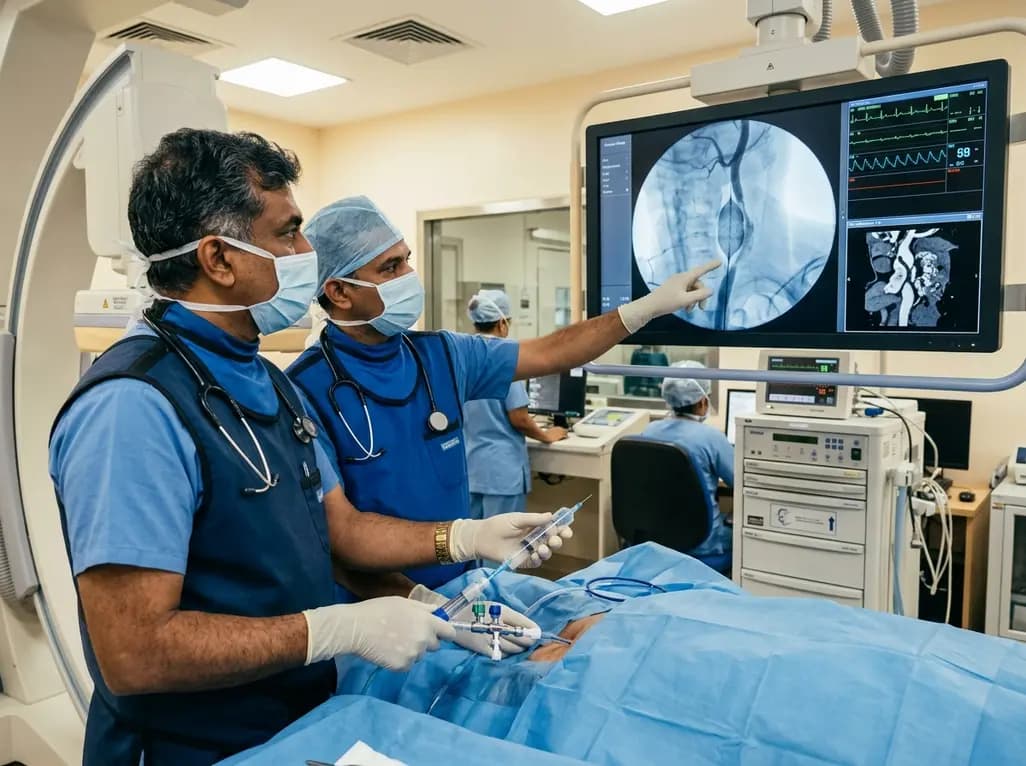
Discover how rescue cutting balloon angioplasty successfully modifies heavily calcified carotid stenosis, enabling stent delivery without baroreceptor instability and ensuring favorable long-term neurologic outcomes.
Today
A breakthrough study reveals that digoxin alleviates osteoarthritis by targeting LSP1 in synovial macrophages, preventing harmful extracellular trap formation and restoring SOD1 activity. This dual-action mechanism protects joint cartilage and offers a promising repurposing strategy for disease modification.
Today

Data from the 2026 Workforce Health Index reveals that 38.7% of urban employees and dependents exhibit abnormal glycemic control. Emerging at increasingly younger ages, this metabolic shift intersects with severe vitamin deficiencies, highlighting the urgent need for early workplace screenings and targeted clinical care.
Today
