.webp&w=384&q=75)

Loading, please wait...
Loading, please wait...
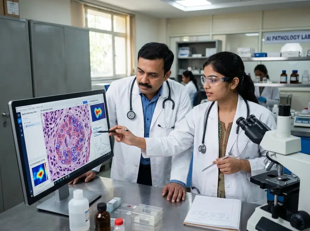
Modern clinical laboratories routinely generate microscopic imaging data that far exceed human analytical capacity. Consequently, pathologists and biomedical researchers face substantial cognitive demands when evaluating complex specimens. Over the past decade, microscopy image analysis has evolved from basic pixel processing into a sophisticated computational discipline powered by artificial intelligence. This paradigm shift enables clinicians to extract reproducible quantitative measurements from cellular structures, neoplastic tissues, and microenvironmental niches. Furthermore, automated computational platforms substantially reduce observer variability across clinical laboratories. As artificial intelligence advances from assisting manual tasks to uncovering novel biological phenotypes, clinicians must understand the mechanisms and practical requirements governing this transformation.
Historically, diagnostic microscopy relied entirely on visual examination of glass slides through conventional optical microscopes. Pathologists evaluated cellular architecture, nuclear pleomorphism, and mitotic figures through manual observation. Although expert human review remains the clinical standard, manual assessment inherently suffers from inter-observer discordance and cognitive fatigue. Therefore, early computational techniques attempted to assist practitioners by applying handcrafted mathematical filters and thresholding algorithms. These deterministic systems quantified staining intensity and detected cell boundaries under strictly controlled conditions. However, conventional algorithms struggled to accommodate technical artifacts, variations in tissue preparation, and biological heterogeneity. As digital slide scanners transformed glass specimens into high-resolution whole slide images, computational demands increased substantially. Deep learning networks subsequently emerged to overcome these technical limitations. Consequently, modern software architectures parse gigapixel histological images within seconds. In addition, these computational frameworks capture subtle morphological features that often escape human perception. By converting qualitative visual observations into standardized quantitative metrics, laboratory medicine gains unprecedented diagnostic consistency. Ultimately, this technological evolution empowers pathologists to focus their expertise on nuanced diagnostic determinations.
Supervised deep learning algorithms established the initial foundation for modern digital diagnostics. Convolutional neural networks learn tissue features from large collections of pathologist-annotated histological slides. For example, supervised models routinely delineate tumor boundaries, quantify tumor-infiltrating lymphocytes, and calculate biomarker expression such as HER2 or Ki-67. Clinicians utilize these quantitative parameters to refine tumor staging and select targeted oncological therapies. Furthermore, supervised segmentation pipelines significantly accelerate screening workflows for infectious agents and hematological malignancies. However, supervised methods depend heavily on exhaustive, expert-generated annotations. Creating these comprehensive datasets requires substantial time and labor from board-certified pathologists. In addition, variations among individual annotators can introduce inadvertent diagnostic biases into trained algorithms. Furthermore, models developed within a single medical center often experience performance degradation when deployed on slides prepared with different stains. Consequently, clinical laboratories must conduct rigorous multicenter validation studies before integrating supervised tools into daily clinical practice. Despite these limitations, supervised algorithms continue to provide immediate operational value in high-volume pathology laboratories.
To circumvent the bottleneck of manual data annotation, researchers have turned to self-supervised learning algorithms. These computational frameworks analyze millions of unlabelled histological images by solving self-generated visual puzzles. Consequently, self-supervised systems learn universal representations of tissue biology without requiring human intervention. Moreover, the emergence of pathology foundation models marks a major leap forward for computational medicine. Rather than constructing narrow tools for individual tasks, researchers pre-train massive models on diverse whole slide libraries across multiple organ systems. Clinicians can subsequently adapt these foundation architectures to specialized diagnostic applications using only small annotated cohorts. For example, fine-tuned foundation models identify occult nodal metastases, subtype difficult sarcomas, and detect rare cellular patterns with high precision. Additionally, generative artificial intelligence models simulate complex tissue microenvironments, helping researchers understand underlying biological mechanisms. Therefore, foundation models transform digital pathology from isolated task automation into a flexible diagnostic platform. However, clinical implementation requires careful oversight to ensure biological plausibility across unique patient demographics.
Clinical deployment of artificial intelligence requires verifiable reliability and complete algorithmic transparency. Standard neural networks frequently generate overconfident predictions, even when presented with corrupted or ambiguous histological samples. Therefore, trustworthy diagnostic platforms must incorporate calibrated uncertainty estimation into their computational core. When an algorithm encounters an atypical histological variant or significant tissue folding, it must quantify its diagnostic confidence. If algorithmic confidence falls below an established threshold, the system automatically triages the slide for manual expert evaluation. This collaborative human-in-the-loop framework prevents erroneous diagnoses in clinical oncology and cytology. Furthermore, software developers must address algorithmic bias resulting from non-representative training datasets. Differences in tissue preparation, staining reagents, and population genetics can degrade performance if models lack broad exposure. Consequently, regulatory guidelines emphasize rigorous external validation on independent, demographically diverse patient populations. Transparent model outputs and calibrated confidence intervals ultimately foster physician confidence and protect patient outcomes during clinical decision-making.
Maximizing the clinical impact of computational microscopy requires dismantling proprietary data silos across healthcare institutions. Historically, vast repositories of digital pathology slides have remained restricted within local institutional databases. To accelerate scientific breakthroughs, academic centers and diagnostic laboratories must adopt interoperable data standards and secure sharing platforms. Furthermore, privacy-preserving techniques like federated learning allow multi-institutional algorithm training without transferring confidential patient records. This collective approach enables researchers to merge whole slide imaging data with spatial transcriptomics, molecular genetics, and longitudinal clinical registries. Consequently, multimodal artificial intelligence models can uncover novel predictive biomarkers for treatment response and disease recurrence. In resource-constrained global health settings, lightweight diagnostic algorithms can assist local clinicians in identifying malaria, leishmaniasis, or tuberculosis from stained smears. Therefore, equitable access to computational microscopy tools can bridge critical healthcare disparities. Sustained collaboration among pathologists, computational biologists, and software engineers will ensure these technologies drive meaningful therapeutic discovery.
Artificial intelligence enhances diagnostic microscopy by automating complex quantitative tasks such as cell segmentation, mitotic counting, and biomarker scoring. By standardizing these measurements across entire gigapixel tissue slides, computational algorithms substantially minimize human subjective error and observer fatigue. Furthermore, calibrated algorithms highlight subtle morphological abnormalities that might otherwise escape routine visual screening, ensuring highly consistent and reproducible diagnostic reporting in clinical pathology.
Foundation models represent large-scale artificial intelligence architectures trained on massive collections of unannotated histology images using self-supervised learning. Consequently, these models capture universal structural and cellular patterns across diverse tissue types without requiring manual human labels. Clinical laboratories can rapidly adapt a single foundation model to diverse specialized tasks, such as detecting rare tumor variants or predicting gene mutations, using very few labeled training samples.
Calibrated uncertainty enables artificial intelligence systems to evaluate their own prediction reliability when processing ambiguous or poor-quality biological specimens. If an algorithm encounters unfamiliar tissue morphology, rare cellular variants, or technical staining artifacts, it calculates an objective uncertainty metric. Rather than delivering an overconfident, potentially inaccurate interpretation, the model immediately flags the slide for pathologist review, safeguarding clinical safety and maintaining diagnostic rigor.
Disclaimer: This content is for informational and educational purposes only and should not be considered as medical advice. Healthcare professionals should make diagnostic and therapeutic decisions based on clinical presentation and individual patient factors. Refer to the latest local and national guidelines for clinical practice.
References

Read summarized clinical updates, watch expert medical content, and earn CME certifications right from your smartphone.
Open your smartphone camera to scan and install.





Artificial intelligence is revolutionizing microscopy image analysis, shifting diagnostic pathology from labor-intensive manual evaluation to quantitative discovery. This article explores how deep learning, self-supervised foundation models, and uncertainty calibration enhance tissue diagnostics.
Today
PZL-26, a selective small-molecule inhibitor of LONP1, reveals essential roles of this mitochondrial protease in complex I biogenesis, translation, and oxidative phosphorylation. These findings provide critical mechanistic insights for developing targeted anticancer therapies against proteostasis-dependent tumors.
Today

A nationwide South Korean study shows underreporting of smoking among women dropped from 57.4% to 36.4% between 2008 and 2021. However, over one-third of female smokers still conceal their habit, highlighting the critical role of cotinine verification in clinical risk assessment.
Today

PepsiCo and Monster Beverage have challenged the FSSAI ban on the energy drink label in Indian courts. While companies highlight supply disruptions, regulatory actions spotlight severe cardiovascular and metabolic risks associated with excessive caffeine, sugar, and taurine consumption, especially in adolescents.
Today
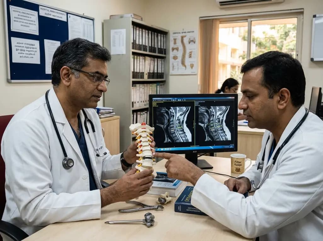
A recent investigation analyzes whether cervical disc arthroplasty effectively restores or merely preserves physiological range of motion, providing critical biomechanical insights for treating hypomobile cervical segments.
Today

A Bayesian multilevel meta-analysis reveals that aerobic training combined with moderate carbohydrate restriction modestly lowers HbA1c in type 2 diabetes. However, sparse data and very low certainty leave incremental benefits over exercise or diet alone unproven, highlighting the need for individualized care.
Yesterday
