.webp&w=384&q=75)

Loading, please wait...
Loading, please wait...
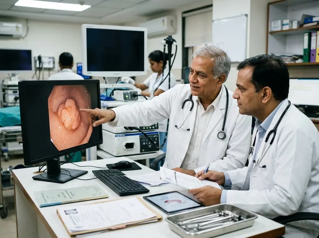
Gastric neoplasms encompass a diverse spectrum of pathological entities, among which type 1 gastric neuroendocrine tumors represent the most frequent subtype. Clinicians frequently encounter these well-differentiated lesions during routine upper endoscopy. While small lesions under ten millimeters typically follow an indolent trajectory, tumors exceeding one centimeter present distinct clinical challenges. Therefore, clinicians must carefully evaluate therapeutic pathways to prevent recurrence while preserving gastric function.
Type 1 gastric neuroendocrine tumors arise primarily in the background of chronic autoimmune atrophic gastritis. Autoimmune destruction of parietal cells leads to severe achlorhydria, which subsequently triggers profound reactive hypergastrinemia by antral G cells. Consequently, chronically elevated serum gastrin exerts a sustained trophic stimulus on enterochromaffin-like cells within the gastric fundus and corpus. Over time, this hormonal stimulation causes sequential hyperplasia, dysplasia, and ultimately multifocal neuroendocrine neoplasia.
Histologically, pathologists classify the vast majority of these lesions as well-differentiated, low-grade G1 or G2 neuroendocrine tumors with low Ki-67 proliferation indices. Despite their favorable biological behavior and excellent overall disease-specific survival, larger lesions carry an increased risk of muscularis propria invasion and regional lymph node metastasis. Furthermore, the persistent hypergastrinemic state creates an enduring field defect throughout the gastric mucosa. As a result, patients remain susceptible to metachronous tumor development even after successful local eradication.
Accurate baseline staging is vital when clinicians detect gastric lesions exceeding one centimeter. High-resolution white-light endoscopy combined with virtual chromoendoscopy allows precise visualization of mucosal architecture and vascular patterns. Additionally, endoscopists should obtain systematic biopsies from both the antrum and the oxyntic mucosa to confirm underlying atrophic gastritis and rule out concomitant adenocarcinoma.
Endoscopic ultrasound plays a pivotal role in staging tumors larger than ten millimeters. Specifically, high-frequency radial or linear echoendoscopes accurately delineate the depth of wall invasion and identify suspicious perigastric lymphadenopathy. Moreover, cross-sectional imaging, including multiphase computed tomography or magnetic resonance imaging, helps exclude distant metastatic spread. In select cases with higher-grade features or suspected extra-gastric involvement, clinicians utilize functional somatostatin receptor positron emission tomography imaging to assess whole-body disease burden before finalizing the therapeutic strategy.
Endoscopic eradication represents the cornerstone of management for localized lesions confined to the mucosa or submucosa. For tumors exceeding one centimeter, endoscopists commonly choose between endoscopic mucosal resection and endoscopic submucosal dissection. While conventional snare resection may suffice for borderline lesions, it frequently leads to piecemeal resection and positive margins in larger tumors.
Conversely, endoscopic submucosal dissection achieves superior en bloc resection rates and histological complete R0 margins. Consequently, this technique facilitates accurate pathological assessment of tumor depth, lymphovascular invasion, and margins. Multiple clinical trials demonstrate that complete endoscopic excision significantly lowers local recurrence rates. However, advanced endoscopic procedures carry inherent risks of intraprocedural bleeding and perforation. Therefore, experienced endoscopists should perform these interventions in tertiary centers equipped with robust surgical backup and comprehensive emergency management protocols.
Although endoscopic therapy remains the preferred primary intervention, selected patients require surgical intervention. Specifically, surgeons consider wedge resection, antrectomy, or partial gastrectomy when tumors exhibit deep muscularis propria invasion, positive margins, or lymph node metastasis. Antrectomy selectively removes the gastrin-producing G-cell mass, thereby normalizing serum gastrin levels and inducing regression of small residual neuroendocrine lesions.
In addition to surgical options, pharmacotherapy provides valuable clinical utility for patients with extensive, unresectable, or rapidly recurrent multifocal disease. Somatostatin analogs, such as octreotide and lanreotide, directly suppress circulating gastrin levels and inhibit enterochromaffin-like cell proliferation through somatostatin receptor subtype 2 activation. Furthermore, novel netazepide formulations targeting gastrin and cholecystokinin-2 receptors offer targeted non-hormonal suppression. Thus, medical therapy serves as an effective organ-preserving bridge or definitive treatment for refractory cases where repeated endoscopic resections become technically impractical.
Multicenter clinical evidence indicates that patients with type 1 lesions larger than one centimeter achieve favorable long-term survival when managed appropriately. Nevertheless, disease recurrence and metachronous tumor emergence remain substantial concerns throughout follow-up. Recent observational cohort studies demonstrate that tumor diameter and incomplete initial resection correlate directly with shorter progression-free survival.
Importantly, composite unfavorable outcomes, including local recurrence, regional lymph node development, and distant progression, predominantly occur within the first three to five years following initial treatment. However, overall disease-specific mortality remains extremely low in well-differentiated cohorts. Multivariate prognostic models highlight that achieving clear histological margins and ensuring thorough surveillance are key determinants of favorable time-to-event outcomes. Therefore, treating teams must balance aggressive tumor removal against the risks of unnecessary radical surgery in this predominantly indolent disease population.
Long-term endoscopic surveillance is essential for all patients following the resection of lesions exceeding one centimeter. Gastroenterologists generally recommend repeat esophagogastroduodenoscopy at six to twelve months post-resection, followed by structured annual or biennial surveillance examinations. During each session, clinicians must meticulously inspect the entire gastric mucosal surface and biopsy any suspicious elevated lesions or prominent mucosal folds.
Furthermore, managing complex cases requires cohesive multidisciplinary collaboration involving gastroenterologists, endocrine oncologists, gastrointestinal pathologists, and visceral surgeons. Multidisciplinary tumor boards ensure individualized treatment plans that integrate patient comorbidities, tumor biology, and procedural risks. By implementing standardized clinical pathways, institutions can achieve optimal oncological clearance while preserving patient quality of life and minimizing intervention-related morbidity.
These tumors develop primarily due to chronic autoimmune atrophic gastritis. The autoimmune destruction of acid-producing parietal cells causes severe achlorhydria. In response, antral G cells continuously secrete high levels of gastrin. This persistent hypergastrinemia exerts a strong trophic stimulus on gastric enterochromaffin-like cells, ultimately inducing hyperplasia, dysplasia, and multifocal neuroendocrine tumors within the stomach lining over extended periods.
Tumors measuring over one centimeter carry a higher risk of invading the submucosa or muscularis propria and metastasizing to regional lymph nodes. Consequently, simple endoscopic surveillance is insufficient. Clinicians must perform endoscopic ultrasound staging and pursue complete en bloc resection via advanced endoscopic submucosal dissection or surgical intervention to ensure clear margins and reduce long-term recurrence risks.
Following successful resection of lesions exceeding one centimeter, guidelines recommend performing a follow-up gastroscopy within six to twelve months. If the stomach remains clear, clinicians continue surveillance every twelve to twenty-four months thereafter. Lifelong endoscopic monitoring remains essential because the underlying autoimmune atrophic gastritis persists, creating an ongoing risk for metachronous neuroendocrine tumors and adenocarcinoma.
Disclaimer: This content is for informational and educational purposes only and should not be considered medical advice. Always consult a qualified healthcare professional regarding any medical condition or before making healthcare decisions. Refer to the latest local and national guidelines for clinical practice.
References

Read summarized clinical updates, watch expert medical content, and earn CME certifications right from your smartphone.
Open your smartphone camera to scan and install.





Discover optimal management strategies, endoscopic resection techniques, and long-term outcomes for type 1 gastric neuroendocrine tumors exceeding 1 cm in diameter, based on multicenter clinical evidence.
last month

A nationwide English cohort study shows that interstitial lung diseases are no longer rare, with incidence jumping 30% and 200,000 active cases. This analysis highlights clinical implications for diagnosis, multi-disciplinary management, and global health priorities across respiratory and internal medicine.
Today

A nationwide South Korean study shows underreporting of smoking among women dropped from 57.4% to 36.4% between 2008 and 2021. However, over one-third of female smokers still conceal their habit, highlighting the critical role of cotinine verification in clinical risk assessment.
Today

PepsiCo and Monster Beverage have challenged the FSSAI ban on the energy drink label in Indian courts. While companies highlight supply disruptions, regulatory actions spotlight severe cardiovascular and metabolic risks associated with excessive caffeine, sugar, and taurine consumption, especially in adolescents.
Today

A new study evaluates the rheological stability of decellularized peripheral nerve matrix hydrogels under varied storage conditions. Findings show -80°C storage and freeze-drying preserve gelation kinetics and mechanical stiffness, offering key insights for shipping and translational clinical applications.
Today

A Bayesian multilevel meta-analysis reveals that aerobic training combined with moderate carbohydrate restriction modestly lowers HbA1c in type 2 diabetes. However, sparse data and very low certainty leave incremental benefits over exercise or diet alone unproven, highlighting the need for individualized care.
Yesterday
