.webp&w=384&q=75)

Loading, please wait...
Loading, please wait...

Orthotopic liver transplantation remains the definitive treatment for patients suffering from end-stage liver disease and acute liver failure. However, perioperative complications frequently compromise patient recovery and allograft longevity. Among these challenges, developing AKI after liver transplantation represents a major clinical hurdle that substantially escalates postoperative morbidity, intensive care stay, and overall mortality. Clinicians historically suspected that subclinical cardiac remodeling contributed significantly to postoperative renal dysfunction. A landmark retrospective study now clarifies this association, examining whether structural left ventricular changes genuinely drive early renal injury in recipients with preserved baseline function.
Cardiovascular remodeling frequently develops in patients with end-stage liver disease due to chronic hyperdynamic circulation and systemic vasodilation. Consequently, clinicians frequently categorize structural cardiac remodeling using two primary echocardiographic parameters: relative wall thickness and left ventricular mass index. Together, these metrics classify ventricular architecture into four distinct phenotypes: normal geometry, concentric remodeling, concentric hypertrophy, and eccentric hypertrophy. In general surgical populations, concentric remodeling and hypertrophy often correlate with impaired coronary flow reserve and perioperative hemodynamic instability. Therefore, investigators hypothesized that pre-existing structural alterations might predispose transplant recipients to renal hypoperfusion during graft reperfusion. However, cirrhotic cardiomyopathy often masks true functional reserve behind elevated baseline cardiac output. To address this question rigorously, researchers analyzed 409 adult deceased-donor liver transplant recipients while strictly excluding individuals with preexisting severe renal dysfunction or overt cardiomyopathy. This specific design allowed investigators to isolate the unique influence of left ventricular geometry on early postoperative renal outcomes.
In this cohort of 409 transplant recipients, early acute kidney injury emerged at an alarming frequency within seven days of surgery. Overall, 189 patients (46.2%) developed renal injury according to standard KDIGO diagnostic criteria. Among these affected individuals, 103 experienced stage 1 injury, 47 progressed to stage 2, and 39 suffered severe stage 3 injury. Interestingly, echocardiographic measurements showed that relative wall thickness, left ventricular mass index, and categorical ventricular geometry were virtually identical between patients who developed renal impairment and those who maintained intact kidney function. Furthermore, multivariable logistic regression analysis demonstrated no significant independent association between abnormal left ventricular geometry and early renal damage. Neither concentric hypertrophy nor eccentric remodeling elevated the odds of postoperative renal failure after controlling for clinical confounders. Thus, structural cardiac alterations alone do not appear to dictate early renal decline in candidates with preserved baseline cardiac function.
Although baseline ventricular geometry showed negligible predictive value, operative and immediate perioperative parameters exerted a decisive impact on renal outcomes. Specifically, extended cold ischemic time demonstrated a statistically significant and independent correlation with the occurrence of renal injury. Prolonged static cold storage intensifies cellular hypoxia, mitochondrial dysfunction, and oxidative stress within the donor organ. Consequently, reperfusion of an allograft subjected to prolonged ischemia triggers massive release of systemic inflammatory mediators and reactive oxygen species. In addition, the volume of intraoperative blood component transfusions independently predicted severe renal impairment. Excessive red blood cell and plasma administration directly reflects surgical hemorrhage, severe portal hypertension, and complex vascular reconstruction. Furthermore, transfusing stored blood products promotes transfusion-related endothelial activation and worsens microvascular renal vasoconstriction. Therefore, surgical precision and shortened preservation times clearly outweigh static cardiac geometry when protecting postoperative renal integrity.
Understanding the interplay between allograft ischemia and systemic renal hemodynamics is essential for modern transplant teams. During the anhepatic phase and subsequent reperfusion, profound hemodynamic fluctuations challenge systemic vascular resistance and cardiac output. When donor livers endure prolonged cold ischemia, post-reperfusion syndrome occurs more frequently and with greater hemodynamic instability. This phenomenon precipitates acute arterial hypotension, elevated central venous pressure, and decreased renal perfusion pressure. Additionally, massive release of damage-associated molecular patterns from the reperfused graft initiates widespread endothelial activation and capillary leak in distant capillary beds. Because the renal medulla operates under baseline hypoxic tension, these systemic surges in inflammatory cytokines induce rapid tubular cell necrosis. Intraoperative fluid shifts and aggressive blood transfusions further heighten renal venous congestion, which impedes glomerular filtration. Consequently, dynamic intraoperative physiological insults, rather than resting cardiac geometry, drive renal tubular damage.
These findings provide actionable clinical guidance for multidisciplinary liver transplant teams, anesthesiologists, and nephrologists. First, clinicians should recognize that preserved ventricular ejection fraction with abnormal geometry does not preclude favorable renal recovery. Instead, transplant teams must concentrate their clinical vigilance on modifiable surgical and logistical variables. Minimizing cold ischemic times through optimized organ transportation, streamlined recipient preparation, and advanced machine perfusion technologies provides substantial renal protection. Moreover, anesthesia teams should implement goal-directed hemodynamic protocols and point-of-care coagulation management using thromboelastography. These targeted interventions effectively reduce unneeded blood product transfusions and prevent fluid overload. Furthermore, close postoperative monitoring of urine output, serum creatinine, and nephrotoxic drug levels allows earlier recognition of renal strain. By addressing operative stressors directly, transplant centers can mitigate complications and improve graft and patient survival.
Cardiologists define left ventricular geometry by calculating left ventricular mass index and relative wall thickness through echocardiography. Relative wall thickness measures posterior wall and septal thickness relative to internal cavity diameter. These measurements divide cardiac structure into normal geometry, concentric remodeling, concentric hypertrophy, and eccentric hypertrophy. These classifications help clinicians detect structural adaptation to chronic pressure or volume overload before major surgery.
Cold ischemic time represents the duration an allograft spends in hypothermic preservation prior to surgical implantation. Prolonged cold storage aggravates cellular ischemia and depletes adenosine triphosphate stores within donor hepatocytes and sinusoidal cells. Upon surgical reperfusion, severely ischemic grafts release substantial pro-inflammatory cytokines and oxygen free radicals into systemic circulation. This inflammatory surge causes systemic vasodilation, acute tubular necrosis, and subsequent kidney dysfunction.
Intraoperative blood transfusion burden indicates severe surgical hemorrhage, coagulopathy, and profound hemodynamic instability during liver transplantation. Transfusing large volumes of packed red blood cells and plasma introduces free hemoglobin, inflammatory mediators, and bioactive lipids into systemic circulation. Consequently, these transfusion components induce endothelial activation, promote renal microvascular vasoconstriction, and heighten oxidative damage within vulnerable renal tubular cells, precipitating post-transplant acute kidney failure.
Disclaimer: This content is for informational and educational purposes only, and does not substitute professional medical advice, diagnosis, or treatment. Refer to the latest local and national guidelines for clinical practice.
References

Read summarized clinical updates, watch expert medical content, and earn CME certifications right from your smartphone.
Open your smartphone camera to scan and install.





A retrospective cohort study of 409 liver transplant recipients reveals that left ventricular geometry does not predict postoperative acute kidney injury. Instead, prolonged cold ischemic time and blood transfusion burden serve as the primary independent determinants of early renal impairment.
last month
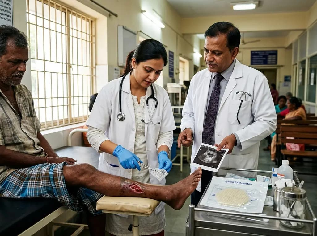
A recent clinical case series highlights the synthetic polylactic acid matrix as an innovative local therapy for refractory pyoderma gangrenosum. The bioresorbable scaffold demonstrated complete ulcer closure and favorable scar quality across chronic wounds, presenting a viable regenerative modality.
Today

A nationwide South Korean study shows underreporting of smoking among women dropped from 57.4% to 36.4% between 2008 and 2021. However, over one-third of female smokers still conceal their habit, highlighting the critical role of cotinine verification in clinical risk assessment.
Today
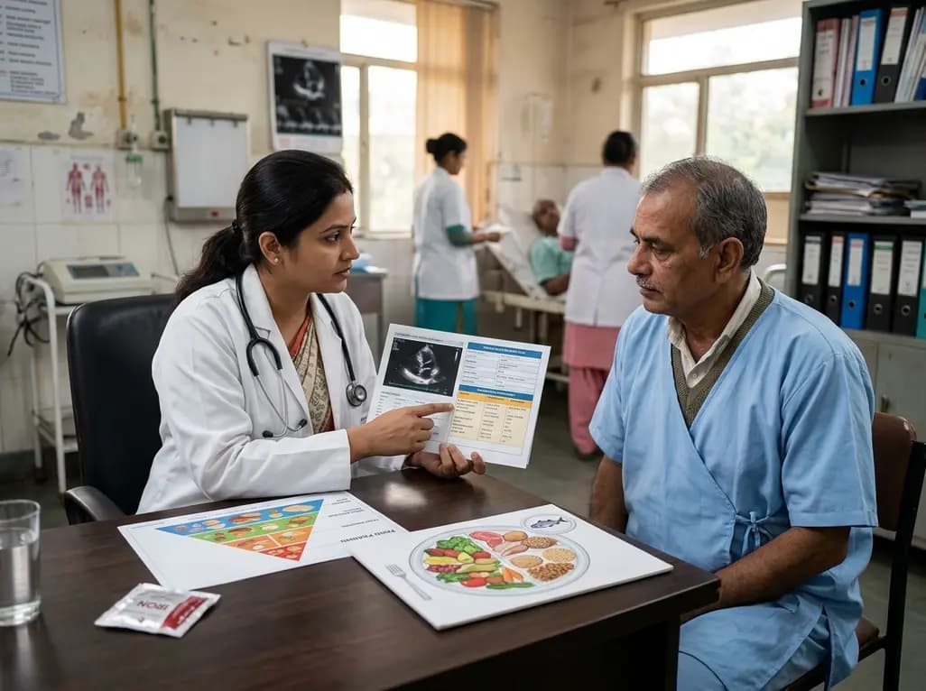
A scientific statement highlights the crucial role of nutrition in managing chronic heart failure. It emphasizes GLIM-based screening, individualized dietary patterns like the Mediterranean diet, appropriate protein and energy intake, avoiding unnecessary fluid restriction, and targeted iron supplementation.
Today

A new study evaluates the rheological stability of decellularized peripheral nerve matrix hydrogels under varied storage conditions. Findings show -80°C storage and freeze-drying preserve gelation kinetics and mechanical stiffness, offering key insights for shipping and translational clinical applications.
Today

A qualitative meta-synthesis highlights the complex interplay between gender dysphoria, minority stress, and eating disorders in transgender and gender-diverse individuals. Clinicians must recognize these distinct drivers to offer supportive, gender-affirming, and interdisciplinary eating disorder care.
Today
