.webp&w=384&q=75)

Loading, please wait...
Loading, please wait...
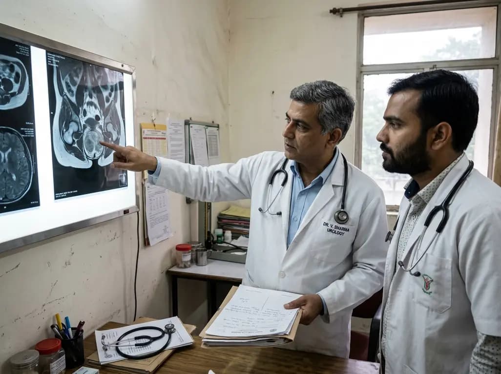
Extrapulmonary manifestations of tuberculosis present significant diagnostic hurdles across global medical practice. Among these rare presentations, testicular tuberculosis represents a challenging condition that frequently mimics testicular neoplasm. In regions with high endemic burdens, healthcare providers frequently encounter atypical presentations of tuberculous infection. However, isolated involvement of the testis without overt pulmonary disease or lower urinary tract involvement remains exceptionally rare. Because scrotal swelling and solid parenchymal masses typically raise immediate suspicion for primary testicular malignancy, clinicians frequently default to aggressive surgical intervention. Consequently, patients with benign infectious lesions often undergo radical inguinal orchiectomy unnecessarily. Furthermore, radiological modalities like standard ultrasonography and contrast-enhanced imaging often fail to clearly distinguish infectious granulomatous processes from malignant testicular germ cell tumors. Establishing early clinical suspicion is essential to preserve fertility, maintain hormonal function, and avoid irreversible surgical interventions. Understanding the subtle clinical nuances and pathology of testicular tuberculosis can significantly aid clinicians in pursuing appropriate diagnostic pathways. Therefore, integrating molecular diagnostics and targeted biopsies into routine urological evaluation forms a critical step toward improving patient outcomes and avoiding unnecessary surgical resection in clinical practice.
A 59-year-old male presented to a tertiary care medical center evaluating a four-day history of progressive, painless left testicular enlargement. Prior to admission, outpatient scrotal ultrasonography identified a solid intra-testicular mass, raising high clinical concern for testicular seminoma. Physical examination revealed an enlarged, firm left testicle with moderate consistency, devoid of significant tenderness or severe systemic symptoms. Diagnostic imaging, including grayscale scrotal ultrasound, contrast-enhanced ultrasound, and enhanced computed tomography, consistently demonstrated features suspicious for primary testicular malignancy. Given the strong radiological suspicion of cancer, the surgical team performed a left radical inguinal orchiectomy. However, postoperative histopathological analysis provided a surprising diagnostic revelation. Microscopic examination of the excised testicular tissue revealed extensive infiltration by acute and chronic inflammatory cells, widespread caseous necrosis, and characteristic epithelioid granulomatous inflammation. These histopathological findings definitively confirmed the diagnosis of isolated testicular tuberculosis. This instructive case highlights how infectious granulomas can seamlessly imitate malignant testicular tumors on conventional imaging, prompting definitive surgical excision before tissue verification. Consequently, clinicians must recognize that rapid onset and ambiguous imaging features warrant a broader differential diagnosis, particularly in high-risk populations where mycobacterial infections remain widespread.
Distinguishing tuberculous orchitis from testicular tumors remains extraordinarily complex due to overlapping clinical and imaging characteristics. Traditional radiological modalities routinely show solid, hypoechoic, or heterogeneous testicular masses in both neoplastic and inflammatory conditions. Consequently, contrast-enhanced ultrasound and pelvic computed tomography scans can misinterpret inflammatory hypervascularity as malignant neovascularization. Moreover, clinical symptoms such as painless testicular enlargement, heavy scrotal sensation, and firm consistency mirror classic presentations of seminomas or non-seminomatous germ cell tumors. Furthermore, routine tumor markers like alpha-fetoprotein, human chorionic gonadotropin, and lactate dehydrogenase often remain completely normal in both isolated testicular tuberculosis and certain seminomas. This overlap creates a misleading diagnostic clinical picture that frequently leads directly to surgical intervention. Furthermore, the absence of active pulmonary tuberculosis or classic constitutional symptoms like fever, night sweats, and weight loss further misdirects clinical judgment. To avoid diagnostic errors, physicians must evaluate subtle history clues, including prior mycobacterial exposures or residence in endemic regions. Additionally, advanced magnetic resonance imaging sequences and contrast dynamics may provide improved soft-tissue characterization. However, definitive differentiation almost always relies on histopathological examination or direct microbiological confirmation. Therefore, relying solely on non-invasive imaging often leads to radical surgical treatments that could otherwise be avoided with prompt medical therapy.
Preventing unnecessary radical orchiectomy requires a structured diagnostic paradigm focused on organ preservation whenever clinical suspicion arises. When clinical findings or imaging characteristics appear atypical, clinicians should strongly consider preoperative or intraoperative frozen section histopathology. Fine-needle aspiration cytology or core needle biopsy can safely yield representative tissue samples without risking tumor spillage if proper surgical protocols are followed. Moreover, incorporating molecular diagnostics such as polymerase chain reaction assays for Mycobacterium tuberculosis enables rapid and highly specific identification of mycobacterial DNA from minimal tissue samples. Furthermore, acid-fast bacilli staining and specialized mycobacterial cultures provide crucial confirmatory evidence while allowing anti-tubercular drug susceptibility testing. Implementing intraoperative frozen section analysis offers immediate guidance during surgical exploration, allowing urologists to switch from radical orchiectomy to organ-sparing management or conservative biopsy. If clinicians identify caseous granulomas or mycobacteria during frozen section analysis, they can avoid testicular removal entirely. Subsequently, initiating standard multi-drug anti-tubercular chemotherapy usually yields full resolution of the infectious lesion while preserving fertility and endocrine function. Therefore, multidisciplinary collaboration between urologists, pathologists, and infectious disease specialists remains paramount in executing organ-preserving strategies for suspicious testicular lesions.
Once healthcare teams establish the diagnosis of testicular tuberculosis, anti-tubercular therapy serves as the primary treatment modality. Standard pharmacological regimens consist of a two-month intensive phase with isoniazid, rifampicin, pyrazinamide, and ethambutol, followed by a continuation phase of isoniazid and rifampicin. This conservative medical approach rapidly resolves local inflammation, reduces granulomatous tissue burden, and eliminates active bacilli. Early pharmacological intervention prevents progressive parenchymal destruction, maintaining Leydig cell function and spermatogenesis whenever organ preservation is achieved. Even in cases where patients undergo orchiectomy prior to definitive diagnosis, postoperative anti-tubercular chemotherapy remains essential to eradicate residual systemic disease and prevent recurrence in the contralateral testicle or extragenital structures. Follow-up protocol requires periodic clinical evaluations and repeat scrotal ultrasonography to confirm complete resolution of inflammatory changes. Moreover, monitoring hepatic and renal function during anti-tubercular therapy ensures patient safety and compliance throughout the extended treatment duration. Public health strategies and clinician education regarding genitourinary tuberculosis further enhance early detection and conservative management across clinical settings. Ultimately, high clinical vigilance combined with accurate pathology testing transforms the management of this rare condition, ensuring favorable therapeutic outcomes and preventing unnecessary organ loss.
Testicular tuberculosis often presents with painless scrotal enlargement, firm testicular consistency, and imaging findings that closely mirror testicular cancer. However, tuberculous lesions may occasionally exhibit localized scrotal skin adherence, subtle epididymal involvement, or sinus tract formation. Because classic constitutional symptoms like fever or weight loss are often missing in isolated disease, clinical differentiation remains difficult without histological or microbiological analysis of tissue samples.
Yes, testicular tuberculosis can be successfully managed without radical orchiectomy if physicians establish an accurate preoperative or intraoperative diagnosis. Standard anti-tubercular chemotherapy effectively eradicates the infection, resolves granulomatous lesions, and preserves fertility and testicular endocrine function. Obtaining tissue via core biopsy or performing intraoperative frozen section analysis allows urologists to confirm the diagnosis safely and avoid unnecessary radical testicular removal.
Preoperative confirmation of testicular tuberculosis relies on tissue biopsy combined with microbiological and molecular testing. Core needle biopsy or fine-needle aspiration provides tissue for histopathology, revealing caseous necrosis and granulomatous inflammation. Additionally, acid-fast bacilli staining, mycobacterial culture, and polymerase chain reaction assays for Mycobacterium tuberculosis DNA offer definitive diagnostic confirmation, guiding appropriate targeted antituberculous therapy while avoiding radical surgical resection.
Disclaimer: This content is for informational and educational purposes only and does not constitute medical advice. Healthcare professionals should evaluate clinical findings independently and refer to the latest local and national guidelines for clinical practice.
References

Read summarized clinical updates, watch expert medical content, and earn CME certifications right from your smartphone.
Open your smartphone camera to scan and install.





Isolated testicular tuberculosis is a rare form of extrapulmonary tuberculosis that frequently mimics testicular malignancy on clinical and imaging evaluations. This comprehensive case review highlights diagnostic pitfalls, key imaging features, and the imperative role of preoperative histopathology.
last month

A multicenter clinical study evaluates fucosylated alpha-1-acid glycoprotein (P-AGP-FR) as an accurate noninvasive fibrosis marker in chronic hepatitis B and C, demonstrating strong correlation with histological staging and superior specificity compared to conventional surrogate indices.
Today

The ClinGen Prenatal Gene Curation Expert Panel evaluated 63 disease relationships across 61 genes, establishing clinical validity for severe fetal phenotypes like hydrops and stillbirth to enhance prenatal genomic interpretation and clinical care.
Yesterday
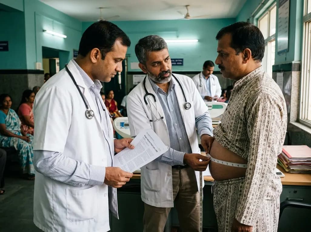
A cohort study demonstrates that the weight-adjusted waist index predicts all-cause and cardiovascular mortality more accurately than BMI and conventional adiposity metrics, offering clinicians a practical anthropometric tool for superior cardiometabolic risk stratification.
Today
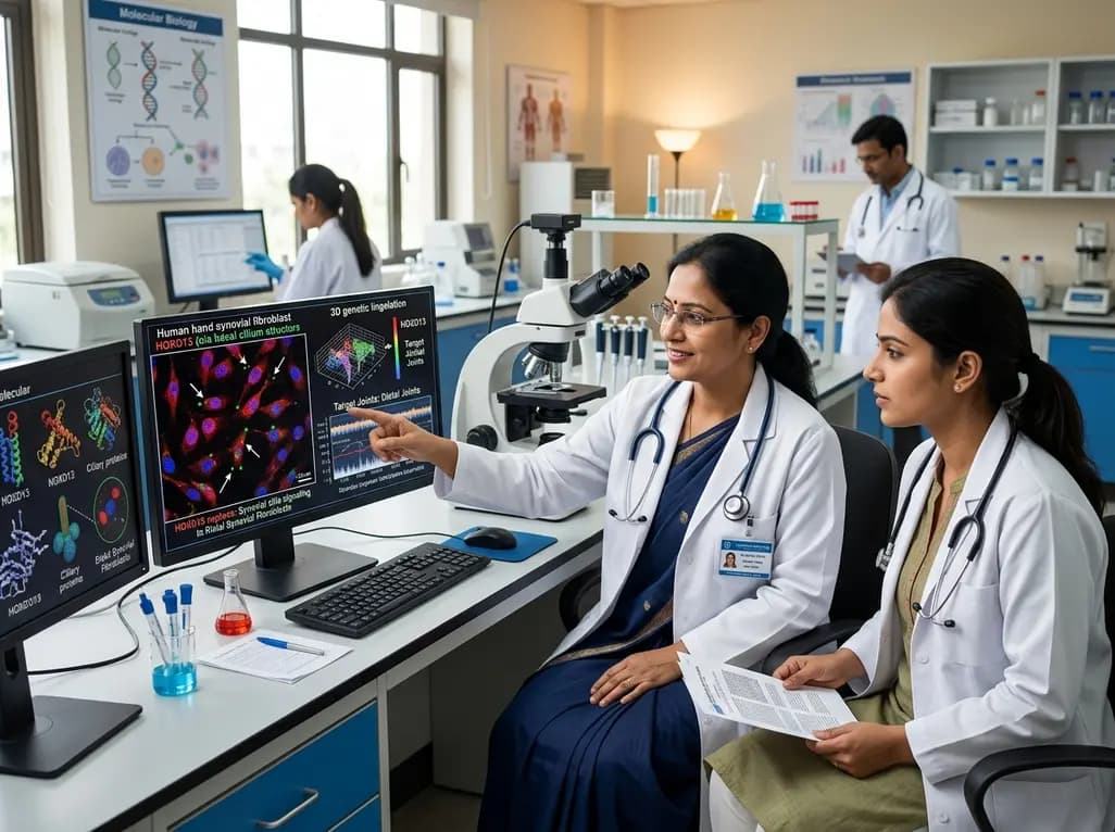
New research reveals that HOXD13 regulates primary cilia signalling and genomic stability in distal synovial fibroblasts, uncovering why conditions like rheumatoid arthritis target specific joints.
Today
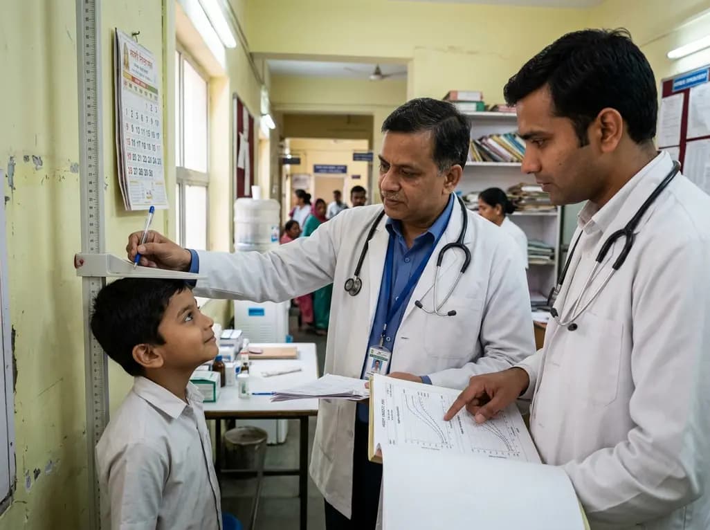
A real-world observational study evaluates pediatric isolated growth hormone deficiency patients transitioning to once-weekly somatrogon, showing sustained height velocity, early IGF-1 shifts, and stabilized BMI.
Today
