.webp&w=384&q=75)

Loading, please wait...
Loading, please wait...
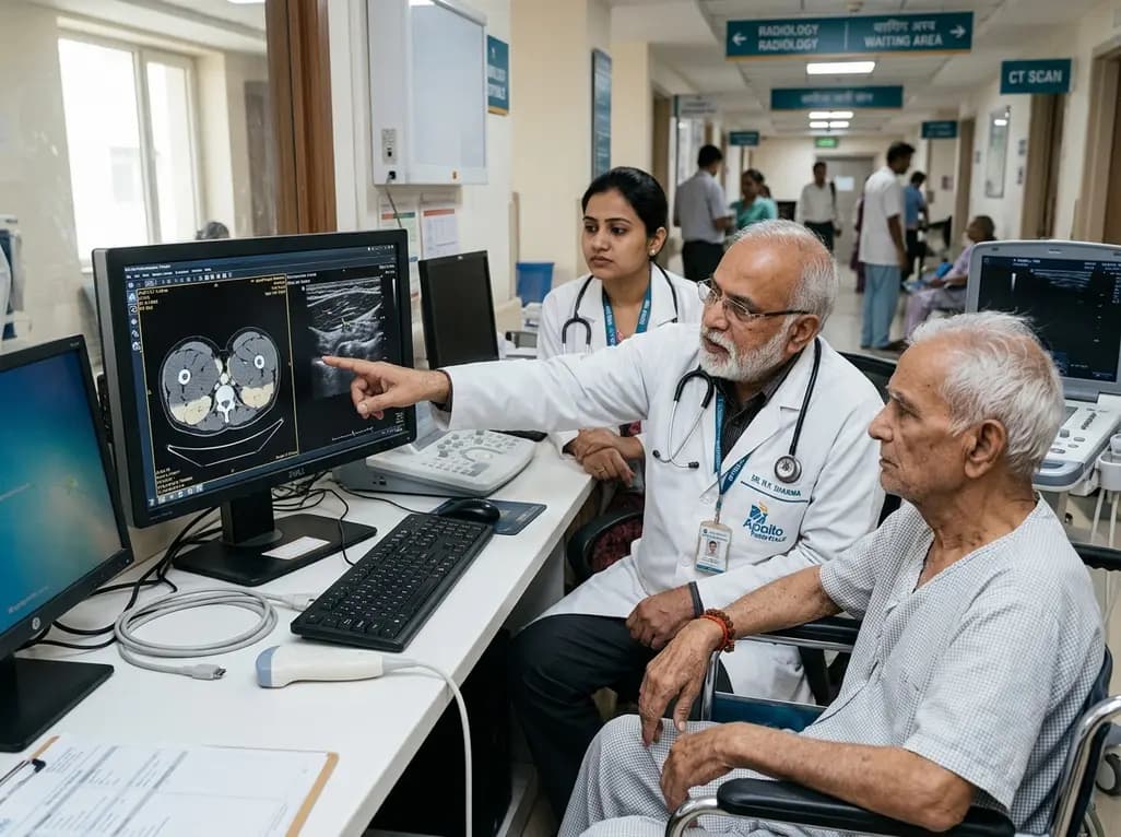
Sarcopenia represents a progressive skeletal muscle disorder characterized by the accelerated loss of muscle mass, quality, and function among aging populations. Clinicians frequently encounter diagnostic dilemmas when identifying early physical decline before significant disability occurs. Traditional functional assessments often provide subjective or effort-dependent metrics that fail to capture underlying structural changes. Consequently, objective imaging muscle assessment has emerged as an essential diagnostic framework in modern geriatric practice. By combining ultrasound architecture and quantitative computed tomography (QCT) tissue composition, clinicians can now quantify structural and functional muscle deterioration with remarkable precision. This comprehensive multimodal approach offers vital clinical insights that empower geriatricians, radiologists, and general physicians to intervene early and improve patient independence.
Age-related muscle degeneration involves distinct pathophysiological pathways that compromise both mechanical force generation and metabolic homeostasis. Standard dual-energy X-ray absorptiometry primarily estimates lean mass but overlooks structural architecture and intramuscular adipose deposition. In contrast, multi-parameter imaging muscle assessment integrates architectural measurements with tissue attenuation values to deliver an accurate physiological profile. Older adults experience muscle fiber atrophy, progressive denervation, and extensive myosteatosis, which degrade physical function long before overt mobility loss manifests.
Furthermore, relying solely on physical performance tests, such as gait speed or grip strength, presents significant limitations in hospitalized or frail individuals. Acute illness, cognitive impairment, or joint pain often confounds physical testing performance. Therefore, objective imaging tools bridge this diagnostic gap by providing quantitative biomarkers independent of patient exertion. By evaluating specific lower-limb muscle groups, clinicians gain actionable prognostic data. This dual architectural and composition evaluation enables earlier identification of sarcopenia risks, facilitating timely rehabilitation protocols and targeted nutritional interventions.
Point-of-care musculoskeletal ultrasound provides a safe, radiation-free, and accessible bedside modality for examining muscle architecture in geriatric patients. Specifically, ultrasound allows precise quantification of the rectus femoris cross-sectional area (CSA), vastus lateralis pennation angle, and gastrocnemius fascicle length. The cross-sectional area of the rectus femoris reflects overall quadriceps mass, which directly correlates with knee extension strength and mobility.
Moreover, architectural parameters like pennation angle and fascicle length reveal the spatial orientation of muscle fibers. A reduced pennation angle indicates severe fiber atrophy and structural disorganization, whereas shortened fascicles diminish the muscle contraction velocity. Research demonstrates that medial gastrocnemius pennation angle and lateral gastrocnemius fascicle length correlate significantly with standardized physical performance batteries. Consequently, sonographic assessment identifies structural vulnerabilities before noticeable clinical weakness occurs. Ultrasound also facilitates serial monitoring during physical therapy programs, providing real-time feedback on muscle hypertrophy and architectural reorganization. Its bedside portability makes it particularly valuable for hospitalized older adults unable to undergo complex radiological examinations.
While ultrasound evaluates superficial muscle architecture, quantitative computed tomography (QCT) offers unmatched volumetric analysis of deep axial musculature and intramuscular fat infiltration. Myosteatosis, the accumulation of inter- and intra-muscular adipose tissue, impairs muscle contraction and promotes systemic metabolic dysfunction. QCT measures tissue attenuation in Hounsfield Units, accurately calculating muscle fat fraction across critical anatomical regions, including thoracic and lumbar vertebrae levels.
Notably, fat fraction within the psoas major muscle at the L4 level serves as a robust independent predictor of reduced gait speed and impaired physical performance. Deep muscular lipid deposition directly undermines biomechanical efficiency and disrupts localized microcirculation. Furthermore, clinicians can opportunistically utilize routine abdominal or pelvic CT scans obtained for other clinical indications, thereby eliminating unnecessary additional radiation exposure. Incorporating QCT-derived fat fractions allows physicians to distinguish genuine muscle tissue from lipid-infiltrated pseudohypertrophy. This compositional detail enhances prognostic accuracy for adverse outcomes, including prolonged hospitalization, recurrent falls, and post-discharge institutionalization.
Combining ultrasound architecture and QCT fat fraction yields synergistic diagnostic power exceeding individual imaging modalities. Recent clinical investigations indicate that cross-modal predictive models incorporating patient age, rectus femoris cross-sectional area, and L4 psoas fat fraction achieve exceptional diagnostic performance. Specifically, integrated models demonstrate an area under the curve (AUC) of 0.912 for detecting decreased gait speed and 0.892 for identifying impaired physical performance.
This cross-modal paradigm successfully captures the multidimensional nature of sarcopenia, bridging the gap between microarchitectural degradation and metabolic fatty replacement. For instance, while sonography captures quadriceps cross-sectional loss, QCT captures core stability degradation through psoas myosteatosis. Consequently, the combined data deliver a comprehensive assessment of overall locomotive capacity. Clinicians can leverage these predictive models to stratify patients into personalized risk categories. High-risk individuals identified through multi-parameter imaging can receive immediate, targeted interventions, mitigating future falls, functional dependency, and secondary complications.
Implementing multi-parameter imaging in routine clinical practice transforms the standard geriatric assessment pathway from reactive management to proactive risk mitigation. Geriatricians and primary care physicians can utilize point-of-care ultrasound during initial consultations to quickly evaluate rectus femoris morphology. When patients undergo cross-sectional spinal or abdominal imaging, automated or semi-automated QCT protocols can simultaneously quantify paraspinal and psoas fat infiltration without increasing scan times.
Furthermore, this multi-parameter framework guides multidisciplinary rehabilitation strategies. Physical therapists can tailor progressive resistance training programs based on specific muscle architecture deficits, such as low pennation angles or reduced fascicle lengths. Similarly, clinical dietitians can prescribe individualized high-protein nutritional supplements and metabolic interventions to target myosteatosis. Additionally, serial imaging assessments allow clinicians to evaluate therapeutic efficacy objectively over time. This systematic approach ensures that therapeutic interventions achieve genuine structural and compositional muscle recovery rather than merely temporary functional compensation.
Technological advancements will continue to enhance the clinical utility of multi-parameter muscle assessment. Artificial intelligence algorithms and deep learning pipelines now automate muscle segmentation and fat fraction quantification on routine CT scans, drastically reducing radiologist workload. Simultaneously, automated ultrasound image analysis is minimizing operator dependency and standardizing measurement repeatability across diverse healthcare settings.
Moreover, emerging modalities like shear wave elastography and contrast-enhanced ultrasound promise deeper insights into muscle stiffness, microvascular perfusion, and extracellular matrix fibrosis. As these technologies integrate into portable handheld ultrasound devices, comprehensive muscle evaluation will become feasible in community clinics, long-term care facilities, and home healthcare settings. Consequently, routine screening will empower clinicians to detect preclinical sarcopenia on a broader scale, revolutionizing preventative geriatric care and healthy aging initiatives worldwide.
Ultrasound evaluates muscle quality by analyzing muscle thickness, cross-sectional area, fascicle length, pennation angle, and echogenicity. Increased echogenicity indicates progressive fatty infiltration and fibrous tissue replacement within the muscle bundle. Concurrently, decreases in pennation angle and fascicle length reflect sarcomere loss and structural disorganization. These architectural alterations directly correlate with reduced contractile force, slower gait velocity, and higher frailty risk.
Quantitative computed tomography fat fraction measurement accurately assesses myosteatosis, which represents the accumulation of intramuscular and intermuscular adipose tissue. Intramuscular fat significantly impairs muscle quality and mechanical efficiency even before substantial muscle mass loss occurs. Identifying elevated fat fractions in axial muscles, such as the psoas major, provides valuable prognostic data regarding gait impairment, fall risks, and metabolic dysfunction.
Cross-modal imaging combines the distinct advantages of ultrasound and quantitative computed tomography. Ultrasound rapidly captures peripheral muscle architecture at the bedside, while computed tomography precisely measures deep axial muscle fat fraction. Integrating these complementary parameters into a unified predictive model yields significantly higher diagnostic accuracy for identifying reduced gait speed and physical impairment than relying on either imaging tool alone.
Disclaimer: This content is for informational and educational purposes only and does not constitute medical advice, diagnosis, or treatment. Healthcare professionals should exercise their independent clinical judgment when evaluating imaging modalities and managing sarcopenia. Refer to the latest local and national guidelines for clinical practice.
References

Read summarized clinical updates, watch expert medical content, and earn CME certifications right from your smartphone.
Open your smartphone camera to scan and install.





A multimodal imaging system combining ultrasound and quantitative CT offers superior diagnostic accuracy for detecting muscle function decline and sarcopenia in older adults.
last month

Chronic eosinophilic pneumonia can present with atypical bronchoalveolar lavage findings and an organizing pneumonia pattern, necessitating surgical lung biopsy.
Today

Discover the crucial distinctions between mesonephric and mesonephric-like proliferations of the female genital tract. Learn the key histomorphologic, immunohistochemical, and molecular differences essential for accurate diagnosis and clinical management.
Today
Kikuchi-Fujimoto disease rarely presents with refractory shock and sinus bradycardia in children. This case report highlights life-threatening hemodynamic instability requiring intensive care, preserved cardiac contractility, and the critical diagnostic role of early excisional lymph node biopsy.
Today

A narrative review investigates whether the articularis genus muscle functions as an independent anatomical entity or blends with the vastus intermedius. We evaluate its morphology, role in retracting the suprapatellar bursa, and direct clinical significance in anterior knee pain and arthroplasty.
Today

A Bayesian multilevel meta-analysis reveals that aerobic training combined with moderate carbohydrate restriction modestly lowers HbA1c in type 2 diabetes. However, sparse data and very low certainty leave incremental benefits over exercise or diet alone unproven, highlighting the need for individualized care.
Today
