.webp&w=384&q=75)

Loading, please wait...
Loading, please wait...

Effective surgical management of drug-resistant epilepsy depends on precise epileptogenic zone delineation. While clinicians typically rely on preictal and ictal stereoelectroencephalography (SEEG) data, interpreting these signals remains challenging. Recent research suggests that examining interictal high-frequency oscillations (HFOs) provides a more reliable complementary approach. Specifically, mapping region-specific HFO rates helps distinguish pathological activity from normal physiological brain signals.
High-frequency oscillations are electrical signals exceeding 80 Hz. Although they are hallmarks of the epileptogenic zone (EZ), they also appear in healthy cortex areas. Consequently, surgeons struggle to identify which HFOs indicate disease. To solve this, researchers retrospectively constructed a normative database from patients who achieved seizure freedom. They compared pathological HFO rates against these normative benchmarks to calculate a specific rate ratio. This method allows for a more objective identification of the seizure-generating tissue.
The study analyzed 63 patients and over 7,500 SEEG recordings to determine the most effective thresholds. Results showed that the HFO rate ratio method accurately localized the EZ in 92% of seizure-free patients. Specifically, the optimal threshold for ripples was 5.8 times the normative value. Furthermore, for fast ripples, the threshold was 2.7 times the normative rate. These thresholds yielded sensitivity and specificity rates exceeding 85%. Moreover, the method identified pathological activity even in patients with persistent seizures, indicating its robust diagnostic potential.
Differentiating between physiological and pathological interictal activity is vital for personalized surgical planning. By using normative benchmarks, clinicians can better predict which brain regions require resection. Therefore, this approach reduces the risk of removing healthy tissue while maximizing the chances of post-operative seizure freedom. Future prospective validation will likely solidify this methodology as a standard in neurosurgical practice.
HFOs are rapid brain signals above 80 Hz recorded during intracranial monitoring. They are divided into ripples (80-250 Hz) and fast ripples (over 250 Hz), acting as biomarkers for the epileptogenic zone.
The ratio compares a patient\'s local HFO rates to a database of healthy cortical activity. If the rate exceeds a specific threshold (e.g., 5.8 for ripples), the area is highly likely to be part of the epileptogenic zone.
Interictal activity occurs between seizures. Analyzing these signals allows clinicians to map the brain without waiting for a seizure to occur, making the diagnostic process faster and potentially safer.
Disclaimer: This content is for informational and educational purposes only. It does not constitute medical advice or establish a doctor-patient relationship. Always seek the advice of a qualified healthcare provider for any medical condition. Refer to the latest local and national guidelines for clinical practice.
References
1. Parasuram H et al. Mapping physiological high-frequency oscillation rates of the cerebral cortex for improved epileptogenic zone delineation in stereoelectroencephalography. J Neurosurg. 2026 Feb 27. doi: 10.3171/2025.9.JNS25602. PMID: 41759173.
2. Frauscher B, et al. High-frequency oscillations: The state of the art and future perspectives. Epilepsia. 2017;58(8):1316-1329.
3. Zijlmans M, et al. High-frequency oscillations as a biomarker for epilepsy. Expert Rev Neurother. 2012;12(10):1231-43.
"
Read summarized clinical updates, watch expert medical content, and earn CME certifications right from your smartphone.
Open your smartphone camera to scan and install.





New research suggests that using HFO rate ratios significantly improves the localization of the epileptogenic zone during epilepsy surgery planning....
7 months ago

Explore why official drug package inserts fail to reflect modern evidence for clozapine prescribing. Learn how legal standards of care, case law, slow titration, and vital adverse event monitoring empower clinicians to prioritize evidence-based medical practice over outdated manufacturer labeling.
Today
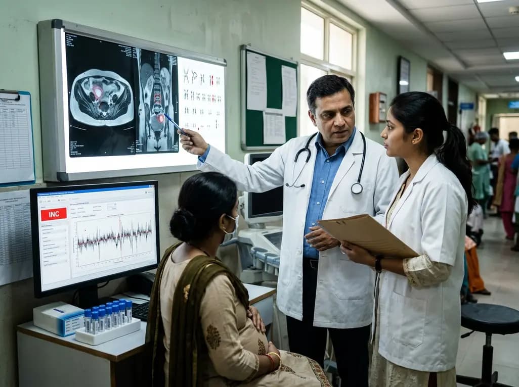
Occult maternal malignancy accounts for 3.82% of persistent unreportable NIPT results with genomic noise. Multiple chromosomal aberrations, notably on chromosome 18, warrant thorough oncological evaluation and whole-body MRI to ensure timely maternal diagnosis while preserving fetal care.
Today

This article reviews the forensic and clinical complexities of acute myocarditis arising on left ventricular hypertrophy in children, highlighting early diagnostic red flags and multidisciplinary postmortem autopsy protocols.
Today
A randomized controlled trial demonstrates that an eight-week dual-task training intervention significantly improves static and dynamic postural control over standard balance exercises in individuals with chronic ankle instability, addressing critical neurocognitive deficits during athletic and daily activities.
Today
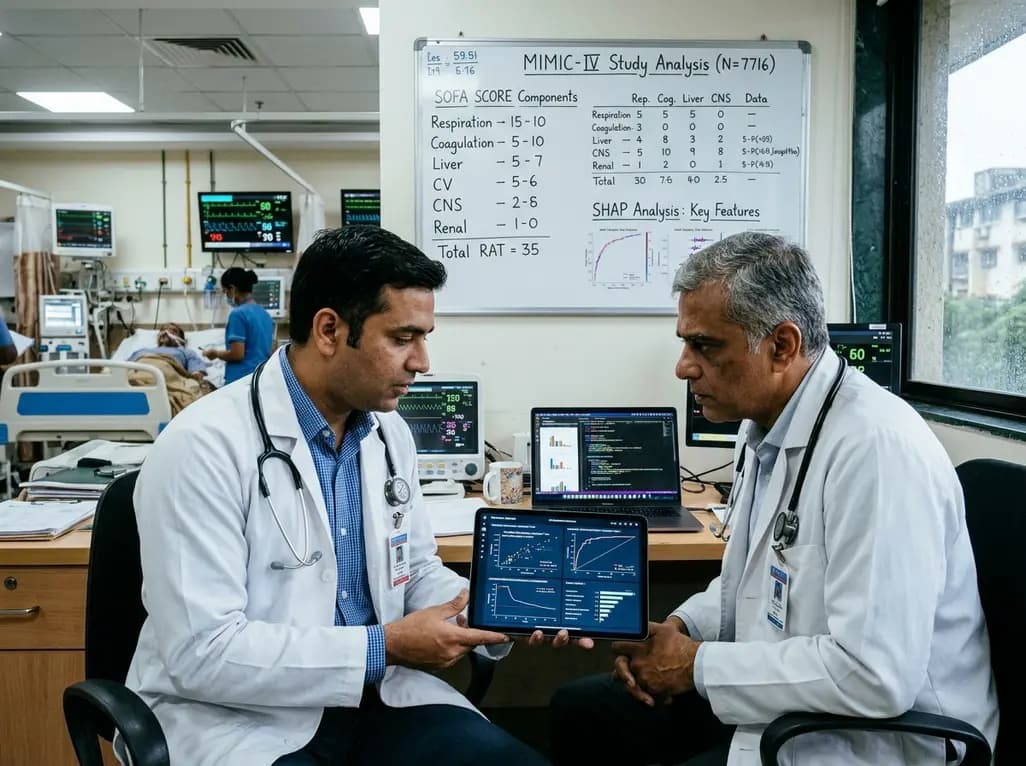
A retrospective cohort study of 7,716 ICU patients from the MIMIC-IV database demonstrates that Gradient Boosting Decision Trees significantly outperform traditional SOFA scores in predicting in-hospital mortality among patients with sepsis and diabetes, with SHAP analysis providing clinical interpretability.
Today
