

Loading, please wait...
Loading, please wait...

Accurate diagnostic assessment of human epidermal growth factor receptor 2 remains a cornerstone in modern oncology. Clinicians routinely depend on reliable HER2 FISH interpretation to guide targeted therapies in patients with breast and gastric malignancies. In standard clinical laboratories, medical laboratory technologists perform the initial signal enumeration, while board-certified pathologists provide the final diagnostic verification. However, laboratories often debate whether pathologists must examine entire fluorescent glass slides or if they can reliably review representative digital images.
Accurate determination of ERBB2 amplification directly dictates therapeutic choices for patients battling invasive carcinomas. For instance, oncologists prescribe targeted monoclonal antibodies and antibody-drug conjugates based on confirmed gene amplification. When immunohistochemistry yields equivocal results, fluorescence in situ hybridization serves as the definitive gold standard. Therefore, clinical laboratories bear an immense responsibility to maintain rigorous precision during enumeration.
However, scoring tumor nuclei presents distinct biological challenges across different tumor types. While breast carcinomas generally present relatively uniform gene amplification, gastric adenocarcinomas display pronounced intratumoral heterogeneity. Consequently, technologists and pathologists must carefully differentiate invasive carcinoma from normal adjacent stroma or carcinoma in situ.
Furthermore, inaccurate signal enumeration risks either denying life-saving targeted treatments or exposing non-amplified patients to costly toxicities. Because therapeutic decisions hinge upon numeric thresholds, laboratories require reproducible scoring protocols. Establishing standardized workflows safeguards clinical accuracy while accelerating turnaround times for critical therapeutic biomarkers across diverse clinical practices.
A recent large-scale retrospective investigation evaluated approximately ten thousand clinical cases across both breast and gastric scoring standards. The primary objective was identifying operational boundaries where technologist enumerations converge reliably with final pathologist interpretations. In typical operational workflows, technologists scan fluorescent hybridization signals, record HER2 and CEP17 counts, and capture representative image fields. Subsequently, pathologists review these findings to issue the definitive clinical report.
Notably, the study revealed that concordance between laboratory technologists and pathologists exceeded ninety-nine percent when signal values fell well outside borderline ranges. When ratios demonstrated clear amplification or marked negativity, pathologist reviews mirrored initial technologist assessments almost perfectly. Thus, extensive data support the clinical validity of initial technologist scoring in distinctly unequivocal specimens.
In contrast, discordant interpretations clustered almost exclusively around official diagnostic cutoffs. When counts hovered near pivotal decision boundaries, minor interpretive variations altered final clinical categorization. These findings demonstrate that concordance depends heavily on numerical distance from established diagnostic cutoffs.
Current diagnostic guidelines define clear cutoffs for both the HER2/CEP17 ratio and average HER2 copy number per cell. For example, breast cancer criteria established by professional societies require a ratio of two or greater, alongside adequate average copy numbers, to confirm gene amplification. However, borderline values frequently generate interpretive challenges during microscopic scoring.
Consequently, cases presenting near these mathematical thresholds require thorough, direct slide examination. Technologists and pathologists may count slightly different fields of view, leading to subtle mathematical discrepancies. In borderline tumors, a shift of a single signal per nucleus can reclassify a patient from negative to equivocal or positive. Therefore, triage algorithms must automatically route borderline cases for comprehensive slide evaluation by a pathologist.
Conversely, tumors with highly amplified signals or unequivocal baseline counts rarely change categories upon secondary review. By defining safe mathematical zones, laboratories can confidently triage cases into rapid image reviews versus full manual slide reassessments. This strategic division protects diagnostic rigor while preserving laboratory resources.
The study analyzed individual technologist performance over extended operational periods to identify factors driving interpretive variation. Interestingly, discordance rates varied among individual technologists independently of overall case volume. High cumulative experience did not automatically guarantee lower discordance rates with pathologist interpretations.
Furthermore, technologist error rates did not consistently decline over successive years of service. This unexpected finding highlights that case volume alone cannot replace ongoing competency evaluations and targeted calibration. Fluorescent signal fading, nuclear overlap, and subjective signal size interpretation introduce subtle individual biases that persist without active feedback.
Therefore, laboratory leaders must institute routine peer reviews and continuous proficiency testing programs. Regular alignment sessions between technologists and supervisory pathologists help standardize scoring habits on challenging cellular morphologies. In addition, implementing structured digital training modules helps align technologist counts with expert pathologist standards, ensuring consistent diagnostic quality across technical staff.
Modern molecular and surgical pathology laboratories handle increasing workloads of diagnostic specimens under strict turnaround constraints. Conducting complete microscopic reassessments on every fluorescent slide consumes substantial pathologist time and creates unnecessary reporting bottlenecks. Consequently, adopting data-driven triaging systems optimizes laboratory workflow efficiency without compromising clinical safety.
Specifically, laboratories can safely direct high-confidence cases—those with concordance rates exceeding ninety-nine percent—to rapid digital image sign-outs. Under this streamlined protocol, the pathologist examines high-resolution captured fields, verifies probe hybridization adequacy, and confirms absence of non-specific background fluorescence. Meanwhile, pathologists reserve comprehensive manual slide reviews for borderline, heterogeneous, or technically ambiguous specimens.
Ultimately, this evidence-based triage model elevates overall patient care by focusing specialist expertise where clinical ambiguity is highest. Laboratories achieve faster report delivery for oncologists while maintaining uncompromising accuracy for targeted therapeutic decisions. Adopting these refined workflows modernizes clinical biomarker testing across global pathology centers.
Breast cancer guidelines evaluate invasive tumor cells using uniform circumferential membrane signals and strict ratio cutoffs. In contrast, gastric adenocarcinoma frequently exhibits marked intratumoral heterogeneity and incomplete basolateral membrane staining. Consequently, scoring gastric specimens demands focused evaluation of cohesive tumor clusters with focal gene amplification. Pathologists must therefore recognize these differing tumor biology patterns when reviewing enumeration data to ensure accurate staging and treatment selection.
Laboratories can safely implement limited review when technologist signal counts sit far outside borderline decision intervals. Specifically, cases displaying extremely elevated HER2/CEP17 ratios or indisputably low baseline counts show concordance exceeding ninety-nine percent with pathologist reviews. Therefore, reviewing digital representative images rather than entire glass slides provides dependable diagnostic accuracy in clearly negative or highly amplified tumors, conserving valuable specialist pathologist time during routine sign-outs.
Tumors near diagnostic thresholds present subtle variations in cellular enumeration and nuclear overlap. Consequently, small counting discrepancies between technologists and pathologists can inadvertently shift clinical classification across positive, equivocal, or negative categories. Comprehensive manual microscopic evaluation ensures proper identification of true invasive tumor areas, eliminates non-neoplastic stromal signals, and prevents catastrophic misclassification in equivocal cases requiring life-prolonging targeted anti-HER2 oncologic therapies.
Disclaimer: This content is for informational and educational purposes only and is not intended as medical advice or as a substitute for professional clinical judgment. Diagnostic thresholds and laboratory workflows should be tailored to individual institutional protocols and validated methodologies. Refer to the latest local and national guidelines for clinical practice.
References

Read summarized clinical updates, watch expert medical content, and earn CME certifications right from your smartphone.
Open your smartphone camera to scan and install.





A retrospective evaluation of ~10,000 HER2 FISH cases reveals >99% technologist-pathologist concordance in unequivocal ranges, supporting efficient sign-out triage in breast and gastric cancer.
Today
A recent investigation reveals that GLP-1 induces IL-27 synthesis in macrophages via the PI3K/AKT signaling cascade. This axis regulates immune polarization and contextual inflammation, offering valuable perspectives for managing obesity-induced metabolic disease and chronic systemic inflammation.
Today
The GendAge weight loss study investigates how sex hormones, age, and menopausal status modulate caloric restriction response. By comparing metabolic adaptations across younger and older cohorts, this controlled feeding trial aims to refine precision dietary guidance for effective obesity management.
Today
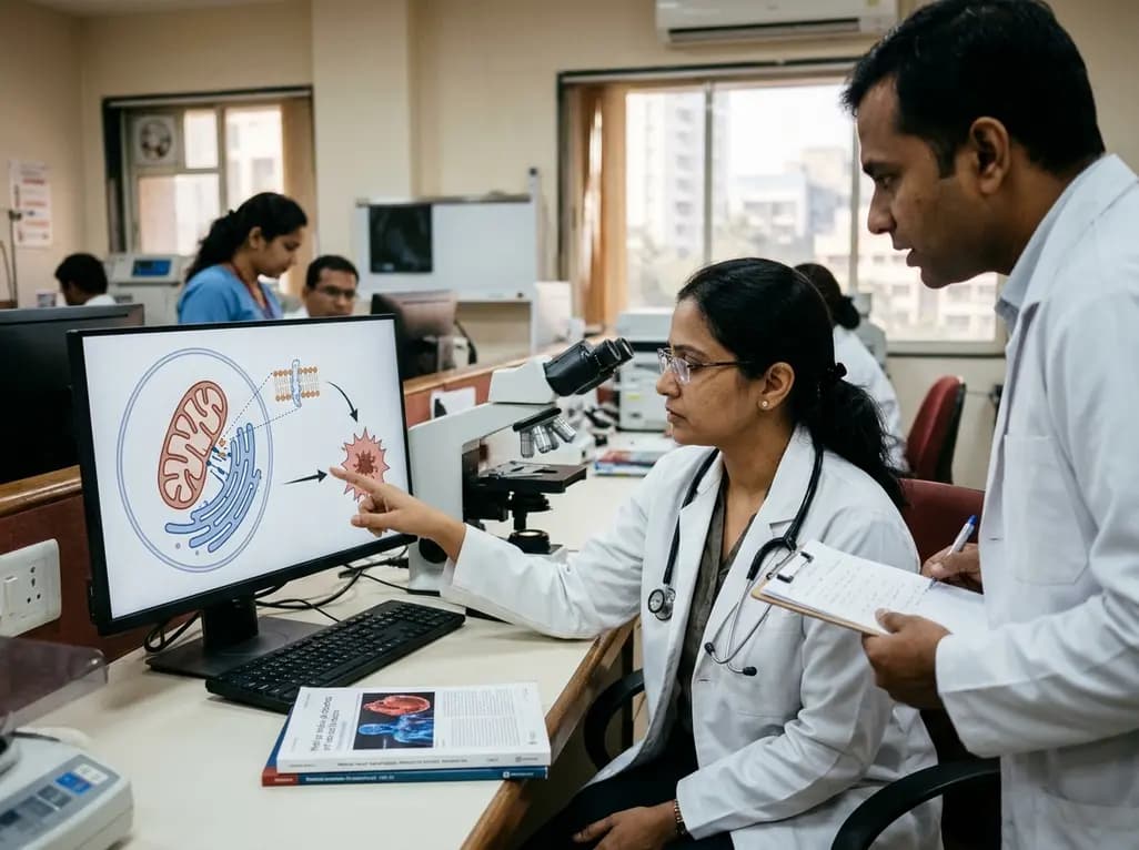
Recent research highlights how endoplasmic reticulum and mitochondrial interactions at contact sites regulate ferroptosis in cardiovascular diseases. This article explores lipid peroxidation, organelle crosstalk, and potential therapeutic strategies for ischemic heart disease and heart failure.
Today
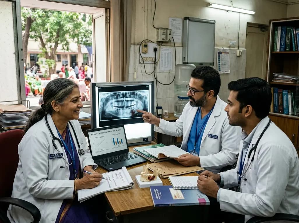
A systematic review demonstrates that teriparatide significantly improves healing in medication-related osteonecrosis of the jaw (MRONJ), especially when combined with surgical and non-surgical therapies. This review explores clinical evidence, biological mechanisms, and treatment protocols for clinicians.
Today
An updated meta-analysis shows that 37% of patients remain euthyroid after stopping thyroid hormone therapy, with success rising to 41% in subclinical hypothyroidism. Clinicians can safely consider supervised deprescribing in selected individuals using structured monitoring.
Today
