.webp&w=384&q=75)

Loading, please wait...
Loading, please wait...
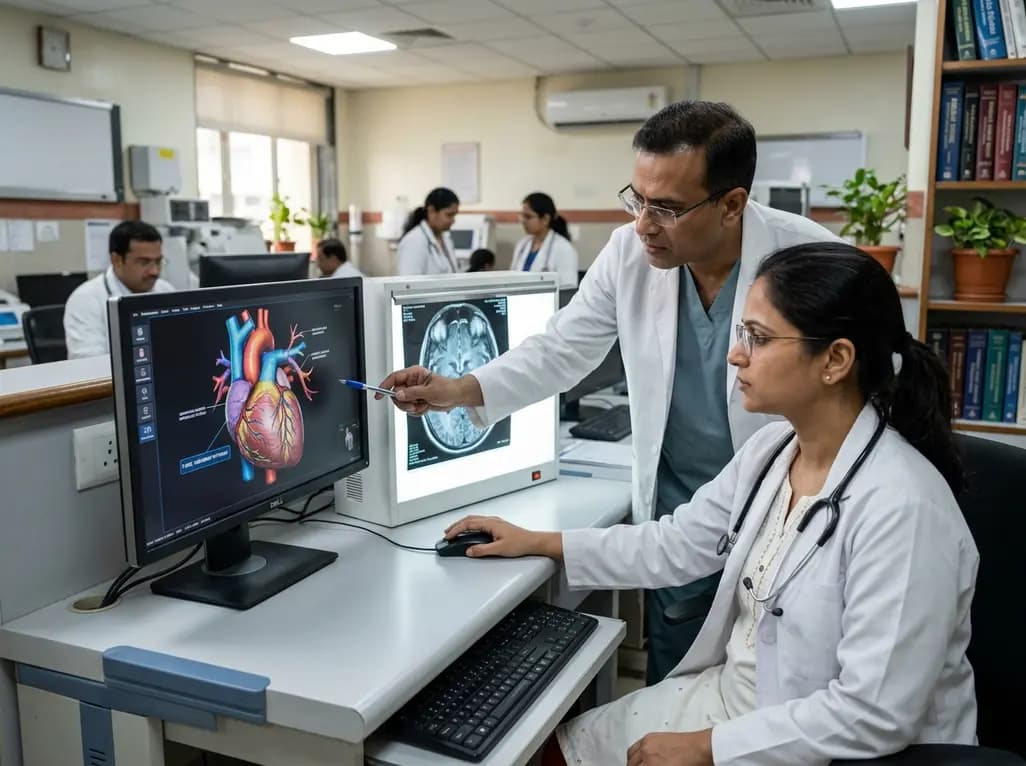
Modern medical education relies heavily on visual clarity, spatial precision, and pedagogical intent. As machine learning advances across healthcare, clinicians and educators increasingly evaluate generative tools for medical visualization. However, recent scientific evaluations emphasize that contemporary models cannot replace trained medical artists. Successfully deploying anatomical illustration AI requires much more than statistical pixel matching. Medical illustrations demand an understanding of physiological relations, tissue planes, vascular variations, and clinical relevance. When software merely combines surface patterns, it frequently produces subtle yet dangerous anatomical inaccuracies. Consequently, relying blindly on automated rendering creates significant risks for surgical education, diagnostic comprehension, and patient counseling. Understanding the genuine capabilities and structural constraints of modern machine learning tools enables medical institutions to integrate novel technologies responsibly without compromising scientific rigor.
Generative algorithms synthesize visual content by analyzing massive training repositories. They detect statistical regularities across millions of biomedical images to reproduce lifelike textures, shading, and colors. Nevertheless, pattern recognition does not equal anatomical comprehension. Human medical illustrators spend years studying regional anatomy, functional mechanics, and surgical approaches. They understand how a fascia layer envelops adjacent muscles, how nerves traverse protective tunnels, and how arteries supply specific tissue beds. In contrast, current algorithms simply predict likely pixel arrangements based on mathematical probabilities. Therefore, the generated graphics often look superficially persuasive while harboring critical structural errors. For instance, a model might illustrate a coronary artery bifurcating into nonexistent branches or place a retroperitoneal structure directly into the peritoneal cavity. Because these algorithms do not understand spatial depth or biomechanical function, they generate plausible fiction rather than anatomical truth. Medical educators must recognize that visual fidelity does not guarantee clinical validity.
Beyond structural distortions, generative systems frequently hallucinate entirely fictitious anatomical features. These hallucinations occur because text-to-image architectures fill ambiguous semantic gaps with speculative pixel arrays. When an algorithm encounters complex prompts involving layered tissue dissections, it attempts to satisfy stylistic constraints rather than biological reality. Consequently, fabricated venous tributaries, distorted neural pathways, or unnatural tissue densities appear in the final rendering. In clinical training, such errors can misguide novice trainees who lack the clinical experience to detect subtle fabrications. Furthermore, artificial intelligence lacks genuine pedagogical intent. An expert medical illustrator deliberately emphasizes specific structures, alters lighting to highlight key landmarks, and simplifies irrelevant clutter to enhance student learning. Generative algorithms cannot exercise this deliberate visual judgment. They treat every pixel with equal algorithmic weight, frequently obscuring vital pathological cues behind extraneous visual noise. Therefore, uncritical adoption of automated imagery undermines didactic efficacy.
Developing a dedicated, functionally aware artificial intelligence system specifically for medical illustration presents substantial economic hurdles. Commercial tech developers naturally direct significant research capital toward large, lucrative markets such as diagnostic radiology, oncology screening, and pharmaceutical discovery. In contrast, medical illustration represents a specialized, highly bespoke niche with limited commercial volume. Training an algorithmic model to achieve flawless anatomical comprehension requires thousands of expertly annotated three-dimensional datasets. Sourcing, standardizing, and verifying these gold-standard datasets demands immense financial and human resources. Furthermore, unresolved legal and copyright issues surrounding proprietary anatomical atlases complicate model training. Tech developers face substantial liability if training pipelines ingest copyrighted artistic works without explicit authorization. Because the addressable market for standalone medical illustration software remains relatively small, private venture capital rarely funds dedicated anatomical generative engines. Therefore, progress in this field cannot rely on isolated consumer software tools.
Fortunately, true anatomical intelligence is developing rapidly within adjacent, high-impact clinical disciplines. In advanced surgical planning, robotic surgery, and personalized medicine, multidisciplinary teams are building sophisticated computational models of human anatomy. These clinical platforms combine high-resolution computed tomography, magnetic resonance imaging, and intraoperative navigation data to construct patient-specific three-dimensional anatomical meshes. Because these models directly guide delicate surgical resections and implant fittings, developers subject them to rigorous validation standards and strict regulatory scrutiny. As computational biomechanics and real-time volumetric rendering advance within surgical oncology and orthopedics, the resulting spatial intelligence will naturally spill over into educational illustration. Rather than building medical illustration software in isolation, future educational platforms will derive their anatomical accuracy from these verified surgical ecosystems. Consequently, future visualization tools will inherit robust biomechanical rules, accurate vascular mappings, and realistic tissue dynamics.
Artificial intelligence will serve as a powerful augmentative tool rather than a replacement for skilled medical illustrators. Professional illustrators bring clinical empathy, communicative purpose, and ethical oversight that algorithms cannot replicate. In future workflows, illustrators can utilize machine learning to accelerate preliminary drafting, texture synthesis, and basic line work. However, human specialists must maintain ultimate editorial authority, meticulously validating spatial relationships, labeling landmarks, and correcting algorithmic hallucinations. Furthermore, developing dependable educational tools requires establishing open-access, peer-reviewed repositories of gold-standard anatomical imagery. Academic medical centers and professional societies must collaborate to create curated datasets that train generative architectures under strict anatomical rules. By combining algorithmic efficiency with rigorous human supervision, the medical education community can safely harness artificial intelligence. Ultimately, maintaining high pedagogical and clinical standards ensures that future visualization technologies enhance student comprehension and safeguard patient care.
Current generative AI architectures rely on statistical pattern matching across vast image datasets rather than true biological comprehension. When prompted with complex medical prompts, these models predict likely pixel sequences to create visually appealing images. Because they do not understand functional relationships, tissue planes, or vascular physiology, they often invent nonexistent blood vessels, misplace organs, or hallucinate aberrant structures to fulfill stylistic parameters.
No, generative AI cannot replace professional medical illustrators. Medical illustration requires deliberate pedagogical intent, deep clinical knowledge, and refined visual judgment to communicate complex surgical concepts effectively. While AI excels at rapid rendering, it lacks ethical awareness, diagnostic context, and the ability to tailor visual clarity for specific educational audiences. Human illustrators remain essential for verifying accuracy and maintaining pedagogical integrity.
Future AI illustration tools will likely achieve anatomical accuracy by leveraging advances in surgical navigation, biomechanical modeling, and personalized medicine. High-precision volumetric models built from verified clinical imaging will provide the spatial rules necessary for accurate rendering. Combined with curated gold-standard training datasets and rigorous human supervision, these systems will transition from basic pattern recognition to genuine, functionally aware anatomical visualization.
Disclaimer: This content is for informational and educational purposes only and should not be considered medical advice. Healthcare professionals should exercise their clinical judgment and refer to official clinical guidelines when making diagnostic or therapeutic decisions. Refer to the latest local and national guidelines for clinical practice.
References

Read summarized clinical updates, watch expert medical content, and earn CME certifications right from your smartphone.
Open your smartphone camera to scan and install.





While generative AI offers unprecedented speed, contemporary models struggle with anatomical accuracy and hallucinations. This analysis explores why medical illustration requires genuine functional comprehension, human supervision, and clinical synergy with surgical planning.
last month

PZL-26, a selective small-molecule inhibitor of LONP1, reveals essential roles of this mitochondrial protease in complex I biogenesis, translation, and oxidative phosphorylation. These findings provide critical mechanistic insights for developing targeted anticancer therapies against proteostasis-dependent tumors.
Today

A nationwide South Korean study shows underreporting of smoking among women dropped from 57.4% to 36.4% between 2008 and 2021. However, over one-third of female smokers still conceal their habit, highlighting the critical role of cotinine verification in clinical risk assessment.
Today

PepsiCo and Monster Beverage have challenged the FSSAI ban on the energy drink label in Indian courts. While companies highlight supply disruptions, regulatory actions spotlight severe cardiovascular and metabolic risks associated with excessive caffeine, sugar, and taurine consumption, especially in adolescents.
Today
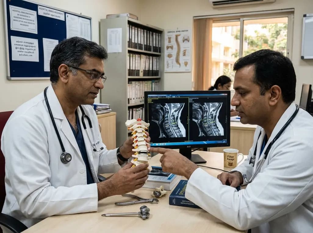
A recent investigation analyzes whether cervical disc arthroplasty effectively restores or merely preserves physiological range of motion, providing critical biomechanical insights for treating hypomobile cervical segments.
Today

A Bayesian multilevel meta-analysis reveals that aerobic training combined with moderate carbohydrate restriction modestly lowers HbA1c in type 2 diabetes. However, sparse data and very low certainty leave incremental benefits over exercise or diet alone unproven, highlighting the need for individualized care.
Yesterday
