.webp&w=384&q=75)

Loading, please wait...
Loading, please wait...

Cerebral palsy represents a leading cause of childhood-onset physical disability worldwide, characterized by non-progressive motor impairments stemming from early brain injury. Evaluating real-time functional neuroimaging in affected children has historically posed substantial logistical challenges. Functional magnetic resonance imaging often requires complete immobility and artificial testing conditions. Consequently, the adoption of fNIRS in cerebral palsy has gained significant momentum across pediatric neurology and neurorehabilitation. Functional near-infrared spectroscopy provides an ecologically valid, non-invasive, and movement-tolerant optical neuroimaging modality. By monitoring relative changes in oxygenated and deoxygenated hemoglobin concentrations, this technology delivers critical insights into hemodynamic responses during active motor execution and resting states.
Functional near-infrared spectroscopy relies on the fundamental principle of neurovascular coupling. When localized cortical neurons fire during a task, local cerebral blood flow increases to fulfill metabolic demands. Optical sensors placed on the scalp emit near-infrared light between 650 and 900 nanometers. Because oxyhemoglobin and deoxyhemoglobin demonstrate distinct light absorption spectra, detectors measure cortical hemodynamic shifts with high temporal resolution. Unlike functional magnetic resonance imaging, fNIRS systems are portable and remarkably resilient to motion artifacts. Therefore, clinicians and neuroscientists can record cortical activity while patients perform functional movements, walk on treadmills, or engage in virtual reality tasks. This ecological validity makes the technology indispensable for pediatric populations who struggle with confined, stationary diagnostic environments.
Recent systematic evidence underscores prominent alterations in sensorimotor cortex activation among individuals with unilateral and bilateral cerebral palsy. During active or passive motor tasks, individuals with cerebral palsy consistently demonstrate a wider spatial extent and higher magnitude of cortical activation compared to typically developing peers. Furthermore, researchers frequently observe atypical ipsilateral hemispheric lateralization during paretic limb movements. This phenomenon indicates extensive cortical reorganization and compensatory recruitment of secondary motor areas. The non-paretic hemisphere often exhibits heightened neural effort to support motor planning and execution. As a result, fNIRS maps these complex bilateral compensatory pathways, providing clinicians with objective topographic data regarding neural reorganization and motor network efficiency.
Beyond sensorimotor regions, fNIRS research highlights the vital role of the prefrontal cortex during motor control. Individuals with cerebral palsy often require elevated cognitive engagement and executive control to execute motor actions that typically developing individuals perform automatically. Studies evaluating dual-task paradigms, virtual reality throwing, and lower-extremity gait tasks reveal pronounced prefrontal hemodynamic responses in this population. The magnitude and pattern of prefrontal activation directly correlate with task complexity and individual functional capacity. Consequently, fNIRS provides a sensitive diagnostic window into cognitive-motor interactions. Tracking prefrontal load helps clinicians design rehabilitation protocols that prevent cognitive fatigue while promoting optimal motor learning.
Investigating functional connectivity through fNIRS offers deeper insights into how disparate brain regions communicate at rest and during activity. Analytical approaches, such as phase-locking values and seed-based correlations, demonstrate altered network topology in children with spastic cerebral palsy. Children with bilateral cerebral palsy often display significantly reduced functional connectivity between the sensorimotor cortex and the somatosensory association cortex. Moreover, resting-state networks show altered clustering coefficients, global efficiency, and interhemispheric coordination. By quantifying these network disruptions, optical imaging uncovers the underlying physiological breakdowns in sensorimotor integration, thereby providing quantitative neural targets for neuromodulatory and physical therapies.
One of the most promising applications of fNIRS involves tracking cortical neuroplasticity following targeted rehabilitation. Longitudinal investigations utilizing robotic-assisted gait training, constraint-induced movement therapy, and virtual reality interventions demonstrate measurable neural shifts. Following effective therapeutic courses, researchers observe a progressive normalization of sensorimotor activation patterns. Specifically, excessive bilateral recruitment diminishes, and neural activation shifts back toward the contralateral hemisphere. Crucially, these neuroimaging changes strongly parallel improvements in validated clinical motor scales. Therefore, fNIRS-derived hemodynamic metrics function as reliable, objective biomarkers of treatment efficacy, enabling clinicians to tailor rehabilitation plans according to individual neuroplastic responses.
Although fNIRS offers extraordinary advantages, several technical considerations warrant careful consideration during clinical implementation. Extracerebral physiological signals, such as scalp blood flow, systemic heart rate, and respiration, can contaminate optical measurements. Implementing short-separation reference channels and advanced filtering algorithms is essential to ensure signal purity. Additionally, standardizing optode placement across diverse pediatric head shapes requires rigorous spatial registration frameworks. Future research must expand sample sizes and explore hyperscanning paradigms to assess social and physical interactions. Integrating fNIRS with electroencephalography and wearable biomechanical sensors will further solidify its role as a cornerstone of individualized precision neurorehabilitation.
Functional near-infrared spectroscopy offers superior portability and high tolerance to patient motion compared to functional magnetic resonance imaging. It allows neuroimaging during upright standing, walking, and natural motor tasks without requiring sedation, auditory isolation, or strict confinement, making it uniquely suited for pediatric patients with cerebral palsy.
Following effective rehabilitation, fNIRS detects a reduction in excessive bilateral cortical overactivation and a return toward contralateral sensorimotor lateralization. These hemodynamic shifts indicate adaptive neuroplasticity, showing that motor planning and execution are becoming more efficient as physical function improves.
Yes, multi-channel fNIRS arrays can simultaneously record hemodynamic activity from both the sensorimotor cortex and the prefrontal cortex. This multi-regional monitoring enables clinicians to evaluate how executive function, cognitive load, and motor execution interact during complex physical tasks or dual-task training.
Disclaimer: This content is for informational and educational purposes only and should not be considered medical advice or relied upon as a substitute for professional healthcare guidance. Refer to the latest local and national guidelines for clinical practice.
References
Khan OA et al. Assessment of cortical activity, functional connectivity, and neuroplasticity in cerebral palsy using functional near-infrared spectroscopy: A scoping review. Dev Med Child Neurol. 2025 Jul. doi: 10.1111/dmcn.16238. PMID: 39963963.
Wang J, Zou Z, Huang H, et al. The functional near infrared spectroscopy applications in children with developmental diseases: a review. Front Neurol. 2025;16:1495138. doi: 10.3389/fneur.2025.1495138.
Huang X, Zhou H, Yang X, et al. Brain functional connectivity and activity during motor control in children with cerebral palsy: a pilot cross-sectional fNIRS study. Transl Pediatr. 2025;14(5):789-799. doi: 10.21037/tp-24-412.

Read summarized clinical updates, watch expert medical content, and earn CME certifications right from your smartphone.
Open your smartphone camera to scan and install.





Discover how functional near-infrared spectroscopy (fNIRS) evaluates cortical activation, functional connectivity, and neuroplasticity in cerebral palsy, providing objective biomarkers to guide neurorehabilitation.
4 weeks back
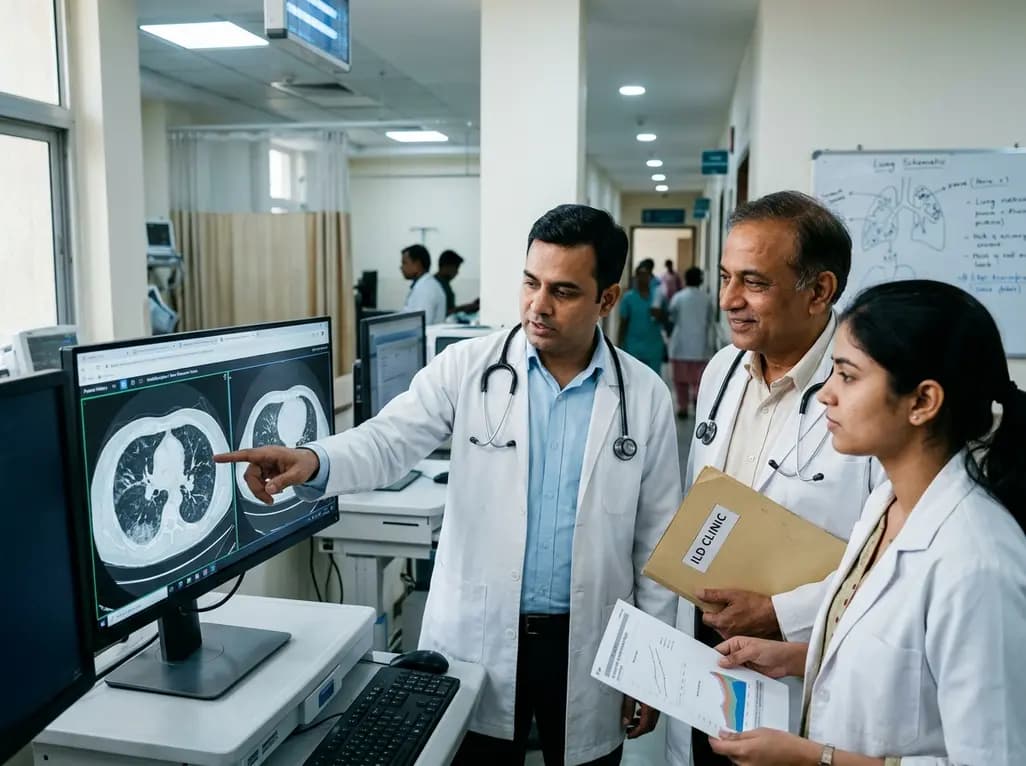
A nationwide English cohort study shows that interstitial lung diseases are no longer rare, with incidence jumping 30% and 200,000 active cases. This analysis highlights clinical implications for diagnosis, multi-disciplinary management, and global health priorities across respiratory and internal medicine.
Today

A nationwide South Korean study shows underreporting of smoking among women dropped from 57.4% to 36.4% between 2008 and 2021. However, over one-third of female smokers still conceal their habit, highlighting the critical role of cotinine verification in clinical risk assessment.
Today

PepsiCo and Monster Beverage have challenged the FSSAI ban on the energy drink label in Indian courts. While companies highlight supply disruptions, regulatory actions spotlight severe cardiovascular and metabolic risks associated with excessive caffeine, sugar, and taurine consumption, especially in adolescents.
Today

A new study evaluates the rheological stability of decellularized peripheral nerve matrix hydrogels under varied storage conditions. Findings show -80°C storage and freeze-drying preserve gelation kinetics and mechanical stiffness, offering key insights for shipping and translational clinical applications.
Today

A qualitative meta-synthesis highlights the complex interplay between gender dysphoria, minority stress, and eating disorders in transgender and gender-diverse individuals. Clinicians must recognize these distinct drivers to offer supportive, gender-affirming, and interdisciplinary eating disorder care.
Today
