

Loading, please wait...
Loading, please wait...

Ultrasound-guided thoracentesis represents an indispensable bedside procedure across modern internal medicine, critical care, and pulmonology units. Clinicians frequently perform this intervention to drain pleural effusions for diagnostic evaluation and therapeutic relief. Ultrasound guidance significantly minimizes the incidence of complications, particularly iatrogenic pneumothorax and visceral organ laceration. Consequently, professional medical societies strongly advise the routine use of point-of-care thoracic ultrasound during needle placement. However, achieving dependable procedural competence requires structured simulation before clinicians perform invasive interventions on real patients. Traditional training phantoms frequently lack anatomical variation and fail to capture the real-time cognitive workload experienced by trainees. Without objective assessment parameters, medical educators struggle to determine whether a learner has truly internalized visual-motor coordination or merely completed manual motions mechanically. Therefore, integrating innovative, metric-driven evaluation platforms into pleural procedural training has become an urgent educational priority.
Simulation educators continually seek robust tools that objectively gauge technical proficiency alongside mental demand. High-fidelity task trainers bridge the crucial gap between didactic knowledge and live patient execution. By standardizing training environments, academic institutions ensure that residents, critical care fellows, and emergency physicians refine their techniques safely. Furthermore, objective measures eliminate subjective evaluator bias, establishing reliable standards for credentialing and clinical readiness.
Eye-tracking technology has emerged as a groundbreaking modality to investigate cognitive ergonomics and user performance in procedural medicine. By capturing optical gaze patterns, wearable eye-tracking glasses record precise visual metrics, such as fixation count, fixation duration, and revisitation frequency. Cognitive load theory suggests that visual attention closely mirrors instantaneous mental processing during complex technical maneuvers. Therefore, analyzing gaze shifts across designated areas of interest illuminates how clinicians distribute visual attention between external anatomical targets and real-time sonographic displays. Excessive fixations or chaotic visual switching often indicate heightened cognitive stress, uncertainty, or divided attention. Conversely, streamlined gaze patterns and concentrated fixations characterize seasoned operators who interpret ultrasound images effortlessly.
In addition, measuring task-evoked optical behaviors overcomes the significant limitations of post-procedure subjective surveys. Traditional psychometric instruments often suffer from recall bias and cannot quantify minute-to-minute fluctuations in mental workload. Wearable eye trackers solve this challenge by generating continuous, non-invasive data streams during live simulation scenarios. Consequently, educators gain deep insights into where clinicians look, how long they process complex visual data, and when cognitive bottlenecks occur. Applying these quantitative metrics directly to invasive pleural procedures establishes rigorous, defensible validity evidence for procedural simulation curriculums.
A recent prospective investigation established preliminary response process validity for an anatomically variable ultrasound-guided thoracentesis phantom using wearable eye-tracking technology. Researchers recruited thirteen experienced clinicians to perform simulated fluid aspiration on a modular phantom engineered with four distinct quadrants of increasing anatomical difficulty. The modular design incorporated varying chest wall thicknesses, rib spacing, and fluid pocket depths to emulate diverse clinical presentations. Investigators equipped participants with eye-tracking glasses to monitor gaze trajectories across two defined areas of interest: the ultrasound monitor and the physical phantom.
The experimental results demonstrated distinct gaze distribution patterns that aligned directly with procedural task demands. Specifically, participants focused their visual attention predominantly on the ultrasound monitor rather than the physical phantom body. The monitor elicited significantly more total fixations than the physical phantom surface, reflecting the intense cognitive processing required for simultaneous sonographic guidance and needle trajectory tracking. Furthermore, the eye-tracking metrics revealed quadrant-specific variations in cognitive workload, confirming that increasing structural difficulty systematically heightened visual processing demands. Post-simulation questionnaires demonstrated high realism ratings, affirming that the phantom faithfully replicated clinical challenges encountered during difficult bedside thoracentesis.
The findings from this simulation trial offer concrete response process validity evidence, confirming that clinician gaze behaviors reflect true task complexity. When operators encounter anatomically complex scenarios, such as narrow intercostal spaces or small loculated fluid collections, their visual gaze patterns shift dynamically. The increased revisitation count between the needle insertion site and the sonographic display illustrates active error-checking and adaptive motor control. Thus, eye-tracking metrics provide an objective physiological window into procedural problem-solving that conventional global rating scales cannot capture.
Importantly, the observed fixation disparity highlights the demanding nature of real-time needle visualization. Clinicians must constantly interpret dynamic sonographic tissue planes while advancing a sharp needle near vulnerable neurovascular bundles. Because the ultrasound display requires intense visual attention, trainees must develop reflexive needle manipulation habits that do not require constant visual monitoring of their hands. Simulation models that capture these visual metrics can therefore identify early procedural flaws before trainees transition to patient care. By pinpointing exactly where visual attention breaks down, clinical instructors can provide targeted coaching to streamline image interpretation and enhance procedural efficiency.
Integrating objective eye-tracking metrics into modular simulation platforms fundamentally reshapes procedural training across emergency medicine, internal medicine, and pulmonology. Modular phantoms that offer graduated difficulty levels enable scaffolded learning pathways. Junior clinicians can begin practice on straightforward anatomical configurations before progressing toward complex anatomy with challenging fluid pockets. This stepped approach prevents cognitive overload during initial training cycles, facilitating superior skill acquisition and long-term retention.
Moreover, objective validity evidence supports standardized competency-based medical education frameworks. Instead of relying strictly on arbitrary procedure logs, training programs can use quantitative gaze efficiency and procedural accuracy to certify independent clinical practice. Clinicians who demonstrate focused gaze control, minimal visual wandering, and steady needle advancement consistently exhibit superior safety profiles at the bedside. Ultimately, implementing validated task trainers with objective workload monitoring optimizes training efficiency, reduces iatrogenic complications, and significantly enhances patient outcomes during invasive bedside interventions.
Eye tracking measures cognitive workload by monitoring physiological gaze metrics, including fixation count, fixation duration, and revisitation rates across designated areas of interest. When clinicians encounter heightened task complexity or uncertainty, their gaze patterns change significantly, showing increased visual fixations and rapid switching between displays. These optical behaviors reliably reflect dynamic mental processing demands and visual-spatial problem solving during intricate interventions.
Ultrasound guidance allows clinicians to visualize the pleural fluid, diaphragm, and underlying lung parenchyma in real time before and during needle entry. This continuous anatomical visualization substantially lowers the risk of accidental organ puncture, hemothorax, and iatrogenic pneumothorax. Consequently, international clinical practice guidelines strongly mandate routine thoracic ultrasound use to maximize patient safety and ensure reliable procedural success.
Modular simulation phantoms provide adjustable difficulty levels, allowing educators to alter anatomical depth, rib spacing, and effusion characteristics. This structural variability supports scaffolded clinical training, guiding learners progressively from simple cases to complex, high-risk interventions. Furthermore, modular task trainers expose trainees to diverse patient anatomies in safe, controlled environments, accelerating technical competence without jeopardizing patient safety.
Disclaimer: This content is for informational and educational purposes only. It is intended to support medical education and clinical learning. Refer to the latest local and national guidelines for clinical practice.
References
Berzaghi BL et al. Gathering Response Process Validity Evidence for an Ultrasound-guided Thoracentesis Phantom Using Eye-tracking Metrics. Simul Healthc. 2026 Nov 09. doi: 10.1097/SIH.0000000000000983. PMID: 43002186.
Anton NE et al. Utilizing Eye Tracking to Assess Medical Student Non-Technical Performance During Scenario-Based Simulation. Glob Surg Educ. 2022;1(1):42. doi: 10.1007/s44186-022-00044-6.
Henneman EA et al. Using Eye Tracking for Measuring Cognitive Workload During Clinical Simulations: Literature Review and Synthesis. Simul Healthc. 2022;17(4):256-264. doi: 10.1097/SIH.0000000000000596.

Read summarized clinical updates, watch expert medical content, and earn CME certifications right from your smartphone.
Open your smartphone camera to scan and install.





A pioneering study demonstrates that eye-tracking metrics provide robust response process validity evidence for an ultrasound-guided thoracentesis phantom, offering objective measurements of cognitive workload to improve procedural simulation and clinical training.
Today
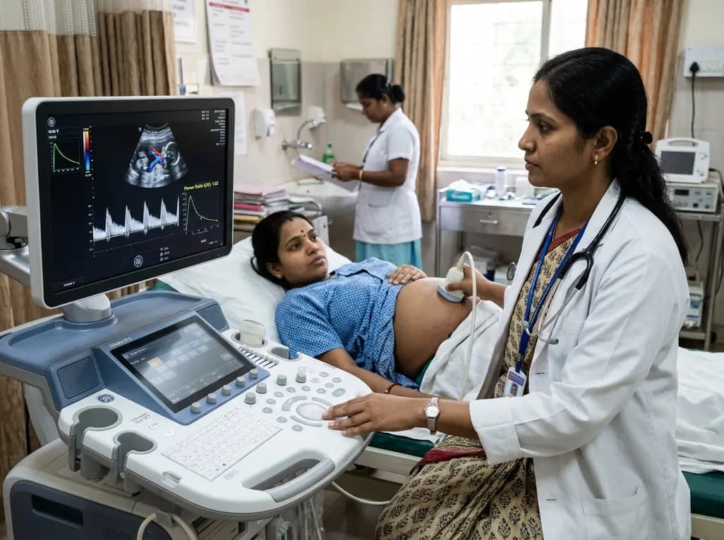
Adaptive spectral analysis of umbilical venous Doppler recordings reveals that an elevated second-trimester maternal-fetal power ratio can distinguish pregnancies at risk of preeclampsia, offering a novel biophysical approach to enhance early vascular risk stratification.
Today

A real-world cohort study of 6,537 patients from the FOUNTAIN platform demonstrates that initiating finerenone significantly reduces albuminuria by 40.7% with minimal eGFR decline and manageable hyperkalemia risk, confirming its practical effectiveness and safety in diabetic kidney disease.
Today

Adding a 12-week tele-supervised resistance band exercise program to intra-articular hyaluronic acid injections significantly improves physical function, chair stands, and mobility in knee osteoarthritis, despite showing no additive reduction in baseline pain scores.
Today

A meta-analysis of 18 RCTs with 77,942 patients reveals that GLP-1 and dual GIP/GLP-1 receptor agonists significantly reduce serum uric acid, cut incident gout flares by 38%, and lower hyperuricemia risk, with dual agonism showing superior urate-lowering efficacy mediated partly by weight loss.
Today
