.webp&w=384&q=75)

Loading, please wait...
Loading, please wait...
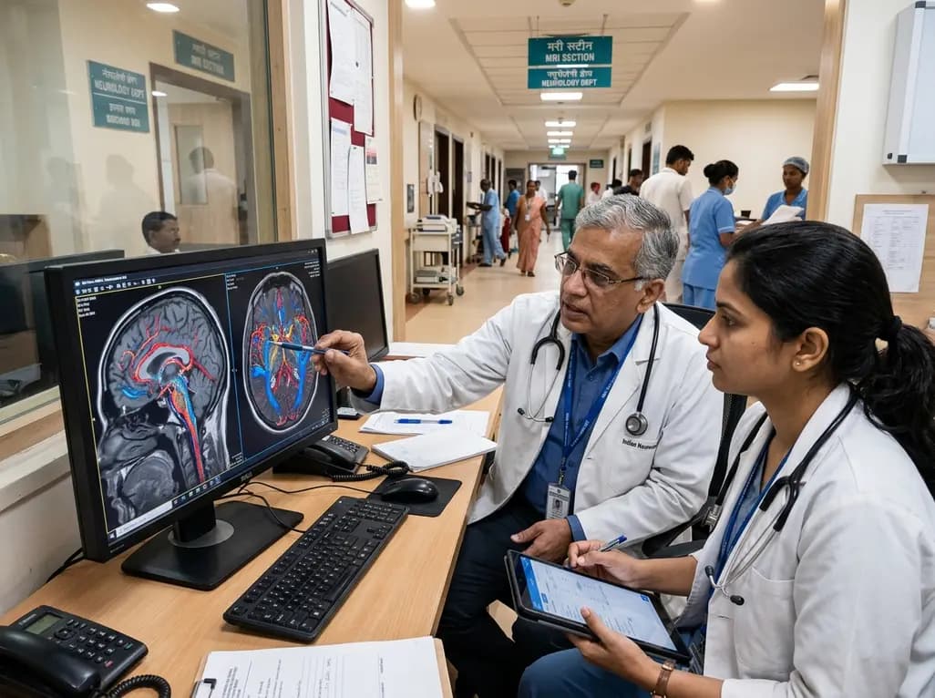
Understanding the physiological mechanisms of cerebrovascular aging remains a cornerstone of modern neurological and cardiovascular research. Normal advancing age leads to progressive structural stiffening of intracranial arteries, higher vascular resistance, and a gradual reduction in resting total cerebral blood flow. These physiological declines frequently correlate with microvascular rarefaction, white matter hyperintensities, and an increased lifetime risk of cognitive impairment. While habitual physical activity is broadly celebrated for promoting overall brain health, the precise alterations within intracranial arterial beds have remained challenging to characterize non-invasively. Advanced neuroimaging modalities, particularly four-dimensional velocity-encoded gradient echo sequence (4D Flow MRI), now provide an unprecedented window into volumetric blood flow, arterial pulsatility, and luminal geometry across the major cerebral conduits.
Age-related vascular remodeling involves complex cellular and extracellular matrix alterations within intracranial conduit vessels, such as the internal carotid arteries and the basilar artery. Consequently, smooth muscle cell senescence, elastin degradation, and increased collagen cross-linking contribute to reduced arterial compliance. This progressive stiffening exposes delicate downstream cerebral microcirculation to excessive kinetic energy and highly pulsatile flow profiles. Therefore, elevated pulsatility index and resistive index often emerge as hallmark indicators of microvascular vulnerability and parenchymal damage. Furthermore, diminishing total cerebral blood flow deprives metabolically active cortical neurons and subcortical pathways of optimal oxygenation and nutrient delivery. Clinicians increasingly recognize that systemic vascular health directly governs brain perfusion, making early identification of hemodynamic disruption critical for preventive neurology.
Traditional non-invasive imaging techniques, including transcranial Doppler ultrasound and standard phase-contrast magnetic resonance angiography, provide limited spatial or single-slice velocity data. In contrast, 3.0-Tesla 4D Flow MRI captures comprehensive, time-resolved three-dimensional blood velocity vectors throughout the entire intracranial vascular tree during a single acquisition. Consequently, researchers can precisely quantify luminal cross-sectional area, net flow rates, and vessel-specific hemodynamic indices across both internal carotid arteries and the basilar artery simultaneously. Because 4D Flow MRI eliminates operator-dependent acoustic window limitations, it yields highly reproducible measures of intracranial arterial mechanics. This cutting-edge quantitative imaging protocol enables detailed evaluations of how long-term lifestyle interventions, particularly endurance running, influence regional vascular resistance and total brain perfusion.
To investigate the relationship between habitual physical activity and cerebral hemodynamics, researchers conducted a prospective cross-sectional study comparing fifty-one long-term endurance runners with fifty-one matched sedentary controls. The athletic cohort maintained at least five hours per week of structured endurance exercise for three or more consecutive years. Using multivariable linear regression and linear mixed-effects models, investigators evaluated bilateral internal carotid arteries and the basilar artery to establish total cerebral blood flow, luminal cross-sectional area, pulsatility index, and resistive index. Consequently, the findings demonstrated distinct structural and physiological adaptations among endurance runners. Long-term aerobic training was associated with favorable luminal dimensions and preserved flow dynamics, highlighting that sustained exercise creates a protective systemic milieu that supports central vascular compliance.
A crucial dimension of the study examined age-by-exercise interactions across young, middle-aged, and older cohorts. While sedentary controls displayed the expected age-dependent elevation in vascular resistance and declining total cerebral perfusion, endurance athletes exhibited significant attenuation of these detrimental trends. Specifically, older runners maintained lower pulsatility and resistive indices in major intracranial arteries compared to their sedentary peers. This hemodynamic preservation suggests that regular aerobic conditioning offsets arterial stiffening, thereby shielding cerebral microvessels from high-pulse pressure trauma. Thus, chronological age alone does not dictate intracranial vascular decline; rather, long-term cardiovascular conditioning serves as a potent modifier of arterial elasticity and brain perfusion throughout the human lifespan.
These findings provide actionable clinical insights for physicians managing aging populations and individuals at elevated cardiovascular risk. Because intracranial vascular stiffening precedes overt cognitive decline, stroke, and vascular dementia, preserving baseline hemodynamic elasticity represents a prime therapeutic objective. Clinicians should actively incorporate structured aerobic exercise prescriptions into primary and secondary preventive care models. Moreover, incorporating advanced non-invasive biomarkers, such as 4D Flow MRI metrics, could enhance risk stratification protocols in clinical trials evaluating neuroprotective interventions. Emphasizing sustained aerobic conditioning throughout midlife and older age may offer a viable non-pharmacological strategy to sustain cerebral autoregulation and maintain lifelong neurological vitality.
Four-dimensional Flow MRI provides comprehensive three-dimensional velocity encoding over the cardiac cycle at high field strength. Unlike conventional two-dimensional phase-contrast magnetic resonance imaging or transcranial Doppler ultrasound, this technique quantifies absolute volumetric flow, arterial pulsatility, resistive index, and luminal geometry across multiple intracranial vessels simultaneously with superior spatial resolution and reproducibility.
Aging causes progressive arterial wall stiffening, loss of elastic fibers, and increased collagen deposition within cerebral arteries. This stiffening increases intracranial pulsatility and downstream microvascular resistance, which transmits harmful kinetic energy directly into fragile cerebral capillaries. Over time, chronic hyperpulsatility impairs cerebral autoregulation, induces white matter ischemia, and accelerates cognitive decline.
In this clinical study, participants engaged in at least five hours per week of structured endurance running for a minimum of three consecutive years. This long-term aerobic regimen was associated with attenuated age-related increases in intracranial arterial resistance, lower vascular pulsatility, and better preservation of total cerebral blood flow across the lifespan.
Disclaimer: This content is for informational and educational purposes only. It is not intended to be a substitute for professional medical advice, diagnosis, or treatment. Always seek the advice of your physician or other qualified health provider with any questions you may have regarding a medical condition. Refer to the latest local and national guidelines for clinical practice.
References
Tang M et al. Intracranial Cerebrovascular Characteristics Associated With Long-Term Endurance Exercise and Age-Related Changes: A 4D Flow MRI Study. J Magn Reson Imaging. 2026 Aug 30. doi: 10.1002/jmri.70520. PMID: 42669028.
Tarumi T, Zhang R. Cerebral blood flow in normal aging adults: cardiovascular determinants, clinical implications, and aerobic fitness. J Neurochem. 2018;144(5):545-555.
Markus HS. Cerebral perfusion and cerebrovascular reactivity in vascular cognitive impairment and dementia. Stroke. 2023;54(2):338-348.

Read summarized clinical updates, watch expert medical content, and earn CME certifications right from your smartphone.
Open your smartphone camera to scan and install.





A cross-sectional 4D Flow MRI study evaluates how long-term endurance exercise influences intracranial hemodynamics, vascular compliance, and total cerebral blood flow across different age groups, offering novel insights into mitigating cerebrovascular aging.
4 weeks back

Abdominal aortic aneurysm rupture remains a catastrophic vascular emergency. Groundbreaking research reveals that USP53 accelerates aneurysm progression by reprogramming smooth muscle cell metabolism toward aerobic glycolysis, highlighting a promising target beyond conventional diameter-based surveillance.
Today

A pharmacometric and machine learning study reveals that specific vaginal bacteria alter cervical antiretroviral exposure, highlighting the need for precision HIV PrEP strategies.
Today

A nationwide Swedish cohort study shows oral glucocorticoid exposure over 1-2 years strongly predicts overall organ damage in SLE, while cardiovascular risk reflects cumulative doses across 3 years. These findings reinforce early steroid minimization and cautious long-term maintenance.
Today

A scoping review highlights neural mobilization as a promising adjunct in managing cervicogenic headache. Integrating neurodynamics into multimodal physical therapy improves pain, range of motion, and posture, though standardized protocols and larger clinical trials are still needed.
Today

Recent research defines ferro-aging as a conserved iron-lipid axis driving organ decline across primates. Age-related iron dyshomeostasis stimulates ACSL4-mediated lipid peroxidation, promoting cellular senescence. Notably, vitamin C directly inhibits ACSL4, suppressing phospholipid oxidation and multi-organ decay.
Today
