.webp&w=384&q=75)

Loading, please wait...
Loading, please wait...

Recent neurophysiological research reveals how the human nervous system connects visual perception with motor execution. Specifically, visually induced kinesthetic illusion refers to the vivid subjective experience of limb movement without voluntary joint displacement. Clinicians in neurological rehabilitation frequently explore virtual reality to stimulate dormant corticospinal circuits. However, whether these illusions directly activate peripheral muscles remains an open question. Now, a pioneering investigation confirms that embodying virtual hands elicits spontaneous peripheral muscle discharges, highlighting significant therapeutic potential.
The human brain constructs limb awareness by integrating visual, proprioceptive, and tactile inputs. Consequently, when clinicians manipulate these sensory streams, they alter bodily self-consciousness. During a kinesthetic illusion, dynamic visual signals override resting proprioceptive feedback from stationary tendons. As a result, the primary motor cortex discharges volleys toward lower spinal circuits. Previous investigations documented these descending cerebral volleys using functional neuroimaging. Nevertheless, researchers struggled to measure consistent corresponding activation in distal target muscles. The concept of embodiment plays a central regulatory role here. Embodiment encompasses three key components: body ownership, self-location, and agency. When patients perceive a virtual hand as their own, the brain processes visual movements as self-generated actions. Therefore, enhancing virtual embodiment strengthens the sensorimotor illusion. Furthermore, stronger illusions amplify central motor outflow to the peripheral neuromuscular junction. Understanding this pathway enables physiotherapists to bridge central motor intention with peripheral muscle recruitment.
To investigate these physiological connections, investigators enrolled twenty-six healthy adult volunteers in an experimental trial. The researchers designed a protocol using video displays of wrist movements while recording electromyography. Specifically, participants observed high-definition recordings of their own hands performing repetitive wrist flexion and extension. The experimental team systematically manipulated two critical factors during these sessions. First, they varied the spatial alignment between the physical hand and the video display. Second, they administered a brief tactile-priming session prior to movement observation. During tactile priming, experimenters brushed the participant's hidden hand while displaying synchronous tactile stimulation on the virtual limb. This tactile synchronization substantially strengthened body ownership ratings among participants. Additionally, participants reported significantly higher illusion intensity during aligned viewing conditions. Through this elegant setup, the team successfully dissociated pure visual observation from true embodied illusion. Consequently, the researchers established a reliable model to assess whether ownership drives motor neuron firing.
The investigative team placed surface electromyography sensors over two primary forearm muscles. Specifically, they monitored the extensor carpi ulnaris and flexor carpi radialis throughout all experimental phases. At first glance, raw condition-level differences in electromyographic amplitude appeared modest. Standard pairwise comparisons failed to show uniform electrical spikes across all test conditions. However, advanced generalized linear mixed model analysis uncovered a profound biological pattern. The statistical modeling revealed that subjective illusion intensity directly predicted peripheral muscle activity. Moreover, tactile priming exhibited a significant positive main effect on sEMG discharge amplitudes. When participants experienced an intense illusion, their peripheral motor units fired spontaneously. In contrast, passive visual observation without embodiment failed to generate meaningful neuromuscular recruitment. Thus, the magnitude of peripheral muscle activation depends directly on perceptual intensity rather than mere visual exposure. These electromyographic findings offer objective confirmation of a functional motor loop linking embodiment to distal effector organs.
These physiological discoveries hold profound implications for modern neurorehabilitation protocols. Following an ischemic stroke or spinal injury, patients frequently experience severe upper-extremity paresis. Traditional active rehabilitation requires baseline motor function, which severely paralyzed individuals lack. In such presentations, passive movement therapies frequently yield limited cortical reorganization. By contrast, visually induced illusions engage frontoparietal motor networks without demanding voluntary muscle contraction. Clinicians can harness embodied virtual reality to bridge this therapeutic void. When medical teams prime paralyzed limbs with tactile cues, they trigger spontaneous motor unit recruitment. Furthermore, this peripheral activity sends afferent feedback toward the sensorimotor cortex, establishing a bidirectional restorative loop. Repeated engagement of these circuits facilitates long-term potentiation and cortical remapping. Therefore, incorporating kinesthetic illusions could accelerate motor recovery in chronic stroke survivors. Ultimately, enhancing embodiment transforms passive observation into an active neuroplastic intervention.
India faces a rapidly growing burden of stroke and traumatic brain injury. Unfortunately, specialized neurorehabilitation centres remain concentrated in metropolitan areas, leaving rural populations underserved. Virtual reality platforms and mobile tablet displays present a scalable solution for this healthcare disparity. Clinicians in Indian tertiary hospitals can readily implement kinesthetic illusion protocols using accessible consumer screens. Furthermore, rehabilitation therapists can easily train community healthcare workers to deliver synchronized tactile priming. Because this technique requires no expensive robotic hardware, public hospitals can deploy it economically. Moreover, home-based tele-rehabilitation programs could incorporate virtual embodiment exercises for chronic hemiparetic patients. Physiatrists can monitor progress remotely while patients perform standardized daily visual-tactile sessions. However, clinicians must establish standardized training protocols to ensure optimal limb alignment. Additionally, clinical teams should consider linguistic adaptations when designing virtual reality user interfaces. Through thoughtful adaptation, Indian healthcare providers can democratize access to advanced neurorestorative therapies.
Ongoing research continues to refine how multisensory feedback optimizes neuroplastic recovery. Emerging digital technologies now allow investigators to pair virtual reality headsets with real-time biometric monitors. Consequently, future clinical protocols could automatically adapt visual movement speeds based on ongoing electromyographic feedback. Moreover, integrating artificial intelligence can help personalize tactile-priming intervals to each patient's sensory thresholds. Indian neuroscientists and biomedical engineers are currently collaborating to develop low-cost virtual rehabilitation suites tailored for resource-constrained environments. These interdisciplinary innovations promise to make immersive therapy widely available outside academic centres. However, large randomized controlled trials in diverse clinical populations remain essential to establish standardized efficacy parameters. Researchers must determine optimal session duration and repetition frequency among hemiplegic individuals. Furthermore, investigators should examine whether combining visual illusions with non-invasive brain stimulation yields synergistic motor recovery. As our mechanistic understanding of embodiment deepens, virtual reality will evolve into an indispensable pillar of modern neurotherapeutics.
Visual observation of movement activates frontoparietal mirror circuits and premotor areas. When individuals perceive virtual limbs as their own, motor cortices generate descending volleys through the corticospinal tract. Consequently, these spinal efferent signals depolarize peripheral motor units, producing measurable electromyographic activity without overt joint displacement.
Synchronous tactile stimulation delivers concurrent visual and somatosensory inputs to the brain. This multisensory integration matches expected sensory feedback with observed actions. As a result, the central nervous system rapidly incorporates the virtual hand into the internal body schema, significantly amplifying both limb ownership and illusion intensity.
Yes, clinicians can effectively apply kinesthetic illusions in flaccid or severely paretic limbs. Because this intervention does not require active volitional movement, it engages dormant sensorimotor pathways safely. Furthermore, combining visual illusion with tactile cues stimulates neuroplastic remapping, preparing paretic pathways for subsequent active functional rehabilitation.
Disclaimer: This content is for informational and educational purposes only... Refer to the latest local and national guidelines for clinical practice.
References

Read summarized clinical updates, watch expert medical content, and earn CME certifications right from your smartphone.
Open your smartphone camera to scan and install.





A pioneering sEMG study reveals that embodied virtual hands intensify kinesthetic illusion and trigger spontaneous peripheral muscle activity. This neurophysiological link provides crucial clinical insights for developing targeted virtual reality rehabilitation protocols in neurological disorders.
Today

Researchers have developed a validated gradient boosting machine learning tool to predict primary tumor origin in cancer of unknown primary using clinical and metastatic variables, achieving high accuracy without initial pathology or genomic testing.
Today
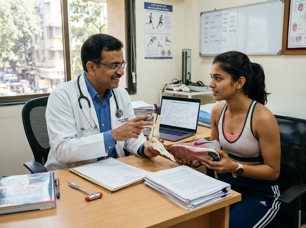
A landmark biomechanical study demonstrates that sports bra support and running shoe cushioning interact significantly to influence tibial acceleration, breast motion, and comfort in female runners, providing crucial insights for clinical counseling in sports medicine and women's health.
Today
A propensity score study demonstrates that thoracotomy-based LVAD implantation preserves postoperative right ventricular systolic function (TAPSE), reduces hospital length of stay by 35%, and lowers long-term mortality hazard compared to standard median sternotomy in advanced heart failure patients.
Today
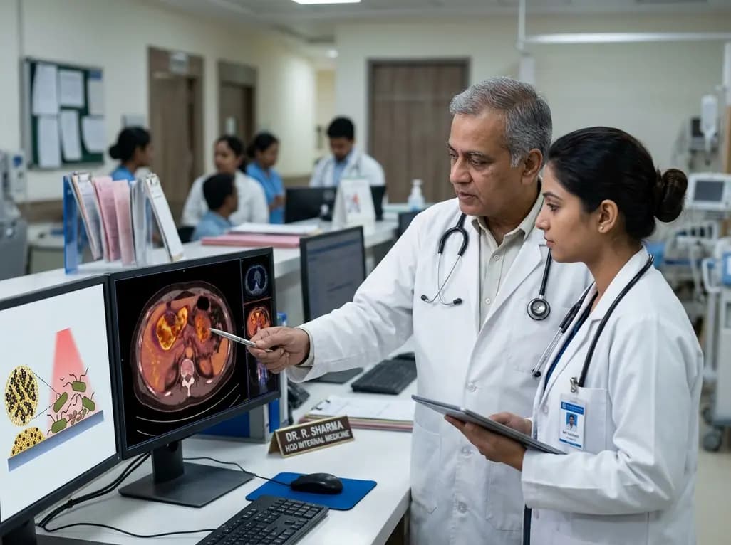
This review explores advances in antibacterial photothermal therapy, highlighting photothermal agents across NIR-I and NIR-II windows. By overcoming biofilm defenses and multidrug resistance without conventional antibiotics, these biomaterial platforms offer transformative options for complex clinical infections.
Today

A landmark Monte Carlo simulation study reveals widespread perchlorate and chlorate exposure through vegetables and drinking water, presenting significant thyroid disruption risks in young children. Learn the physiological mechanisms, dietary drivers, and preventive strategies for clinical practice.
Today
