.webp&w=384&q=75)

Loading, please wait...
Loading, please wait...
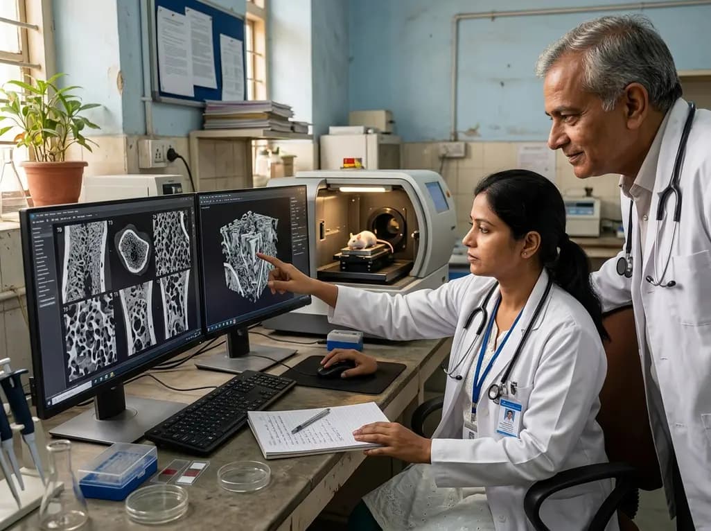
Over the past decade, bone research has significantly shifted from evaluating total areal mineral density to analyzing intricate microstructural architecture. Researchers now recognize that cortical bone porosity represents a key determinant of skeletal strength and fracture risk. Early diagnostic tools focused primarily on trabecular volume, but recent advancements highlight the critical role of cortical thinning and intracortical pore expansion in metabolic bone diseases. Consequently, scientific interest in dynamic tracking techniques has grown rapidly. In preclinical models, monitoring structural changes over time allows investigators to observe early physiological adaptations before macro-level bone loss occurs. Furthermore, understanding these subtle structural variations helps clinicians comprehend how systemic factors compromise skeletal integrity. By evaluating cortical porosity across various physiological states, researchers gain invaluable insights into metabolic bone disorders like osteoporosis. Therefore, modern imaging paradigms emphasize longitudinal observation, establishing a foundation for evaluating novel therapeutic strategies that preserve bone quality.
Preclinical skeletal assessment previously relied on static, destructive histomorphometry to evaluate bone turnover. Although histology provides detailed cellular snapshots, it prevents sequential observation within the same living subject. Fortunately, high-resolution micro-computed tomography has revolutionized this field by providing non-invasive volumetric measurements over extended periods. Longitudinal time-lapse imaging allows researchers to track individual basic multicellular units throughout their operational lifecycle. By registering sequential three-dimensional datasets, scientists can quantify precise localized rates of bone formation and resorption. Consequently, this temporal capability eliminates inter-animal variability, which drastically reduces the sample sizes required for rigorous experimental protocols. In addition, time-lapse protocols provide unprecedented temporal clarity regarding structural remodeling kinetics under varying mechanical or metabolic stresses. As imaging technology continues to improve in spatial resolution, researchers can detect subtle architectural shifts that previously remained undetected during conventional end-point analyses.
While laboratory micro-CT scanners excel at imaging trabecular architecture, capturing minute cortical pores presents distinct technical challenges. To overcome resolution limitations, scientists developed synchrotron propagation phase-contrast micro-computed tomography. This advanced modality leverages highly coherent X-ray beams to produce superior soft-tissue contrast and sub-micron spatial resolution. Consequently, synchrotron imaging enables dynamic visualization of individual cortical remodeling spaces in larger animal models such as rabbits. Researchers can now observe osteoclastic resorption canals expanding and filling in real time with remarkable clarity. Furthermore, this technique captures subtle density variations across newly mineralized osteons, revealing how systemic therapies modulate tissue density. However, synchrotron facilities require specialized infrastructure, which limits widespread accessibility for routine laboratory screening. Despite these logistical constraints, synchrotron phase-contrast imaging remains the gold standard for validating preclinical remodeling models and benchmarking commercial micro-computed tomography systems.
Time-lapse micro-CT imaging provides quantitative metrics that refine classical histomorphometric principles. Historically, researchers inferred linear erosion rates inside resorption cavities through indirect static measurements in canine models. Modern dynamic imaging directly validates these historical estimations by tracking active osteoclastic fronts in living tissue. Studies demonstrate that linear erosion rates remain remarkably consistent across species under physiological baseline conditions. However, pathobiology or pharmacological interventions alter these kinetic profiles significantly. For instance, accelerated or irregular erosion rates alter local pore geometry, which severely compromises structural mechanical integrity. In addition, dynamic voxel-tracking algorithms enable precise calculation of directional mineralization fronts. By measuring active surface areas and volume shifts, researchers can distinguish between balanced coupled remodeling and uncoupled pathological resorption. Consequently, these dynamic metrics provide vital mechanistic benchmarks for evaluating bone fragile states in metabolic conditions.
Pharmacological agents profoundly alter cortical pore dynamics, as demonstrated through longitudinal time-lapse imaging studies. Parathyroid hormone therapies trigger complex remodeling space morphologies while simultaneously reducing localized linear erosion rates. This counterintuitive shift suggests that parathyroid hormone expands overall active remodeling surfaces while moderating individual osteoclast progression speed. Consequently, parathyroid hormone dosing induces unique, highly branched pore networks within compact cortical bone. Conversely, glucocorticoid administration disrupts linear advance patterns altogether, causing chaotic and incomplete osteonal formation. Glucocorticoids suppress osteoblast differentiation and longevity, which leaves resorption cavities unfilled for prolonged periods. Furthermore, dynamic imaging reveals how glucocorticoid exposure increases endosteal resorption and expands intracortical void volume. Understanding these contrasting pharmacological signatures allows endocrinologists and orthopedic researchers to refine drug regimens. Ultimately, dynamic monitoring helps optimize anabolic therapies while mitigating catabolic bone destruction in clinical settings.
Translating preclinical time-lapse imaging findings into clinical practice holds immense promise for patient care in India and globally. High-resolution peripheral quantitative computed tomography currently allows clinicians to monitor human peripheral cortical bone structure in vivo. As image processing algorithms improve, clinical modalities will incorporate insights gained from preclinical synchrotron and time-lapse micro-CT studies. Consequently, physicians will better identify early cortical degradation in patients suffering from postmenopausal osteoporosis, secondary hyperparathyroidism, or chronic kidney disease. In addition, dynamic imaging models provide crucial safety and efficacy data for novel osteoanabolic and antiresorptive agents before human clinical trials. Furthermore, integrating dynamic bone quality metrics into risk assessment models will enhance personalized osteoporosis management. Ultimately, continuous advances in dynamic cortical imaging promise to bridge basic bone biology with improved clinical fracture prevention strategies worldwide.
Monitoring cortical bone porosity is essential because cortical bone provides primary structural strength against mechanical loading and fracture risks. While trabecular bone loss occurs rapidly, age-related and metabolic cortical pore expansion contributes significantly to long-term skeletal fragility. Advanced longitudinal imaging allows researchers and clinicians to identify subtle intracortical structural deterioration early. Consequently, tracking these microstructural changes helps evaluate underlying disease progression and guide targeted therapeutic interventions effectively.
Traditional bone histology requires destructive tissue sectioning, which restricts analysis to a single static time point per animal. In contrast, time-lapse micro-computed tomography non-invasively scans living models at multiple sequential time points. This longitudinal approach allows direct visualization of spatial remodeling dynamics and basic multicellular unit progression over time. Furthermore, time-lapse imaging eliminates inter-subject anatomical variability, significantly reducing experimental sample sizes while increasing statistical power.
Glucocorticoids severely impair cortical bone remodeling by disrupting the coordinated linear advance of mineralization fronts. Pharmacological exposure suppresses osteoblast activity and accelerates osteocyte apoptosis, which leaves osteoclastic resorption pits unfilled for extended durations. Dynamic time-lapse imaging demonstrates that glucocorticoids increase overall cortical porosity and void coalescence. Consequently, these structural disruptions weaken the cortical shell, leading to elevated clinical fracture risks in treated patients.
Disclaimer: This content is for informational and educational purposes only and does not constitute medical advice, diagnosis, or treatment. Healthcare professionals should rely on their professional clinical judgment and refer to the latest local and national guidelines for clinical practice.
References

Read summarized clinical updates, watch expert medical content, and earn CME certifications right from your smartphone.
Open your smartphone camera to scan and install.





A ten-year review highlights how preclinical time-lapse micro-CT and synchrotron imaging allow dynamic tracking of cortical bone porosity and remodeling space kinetics. Recent studies reveal how therapeutic agents like PTH and glucocorticoids alter linear erosion rates and pore morphology.
last month

A comprehensive validation study using PPMI cohort data establishes the psychometric reliability, factor structure, and minimal detectable change of the SCOPA-AUT scale across prodromal and established Parkinson's disease, highlighting its longitudinal association with non-motor daily living experiences.
Today

The ClinGen Prenatal Gene Curation Expert Panel evaluated 63 disease relationships across 61 genes, establishing clinical validity for severe fetal phenotypes like hydrops and stillbirth to enhance prenatal genomic interpretation and clinical care.
Today

A cohort study demonstrates that the weight-adjusted waist index predicts all-cause and cardiovascular mortality more accurately than BMI and conventional adiposity metrics, offering clinicians a practical anthropometric tool for superior cardiometabolic risk stratification.
Today

New research reveals that HOXD13 regulates primary cilia signalling and genomic stability in distal synovial fibroblasts, uncovering why conditions like rheumatoid arthritis target specific joints.
Today

A western India study demonstrates that shortened coagulation times (PT, APTT), elevated mean platelet volume, and increased UACR strongly correlate with type 2 diabetes microvascular complications, highlighting the clinical value of combined hemostatic and renal profiling for early risk stratification.
Today
