.webp&w=384&q=75)

Loading, please wait...
Loading, please wait...
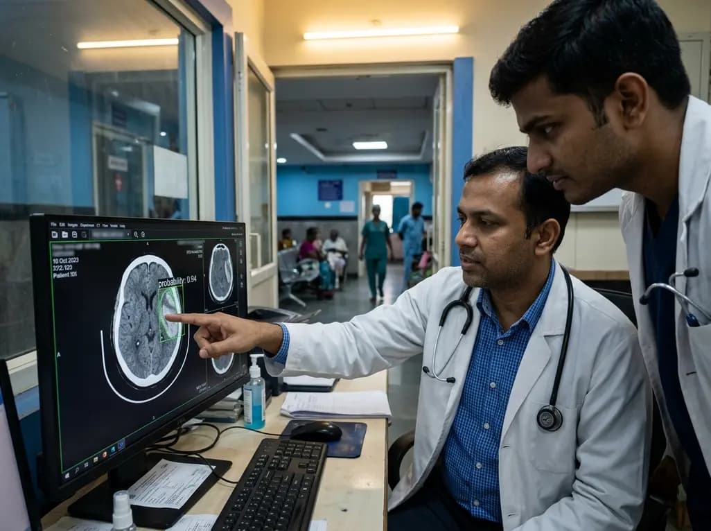
Accurate and rapid evaluation of head computed tomography scans remains a critical step in emergency neurological care. Non-contrast computed tomography scans are the primary imaging tool used when evaluating acute head trauma or sudden neurological decline. However, manual interpretation of these scans can be time-consuming, subjective, and prone to error during high-volume shifts. Consequently, deep learning solutions have gained substantial traction to assist radiologists and clinicians. Automated subdural hematoma detection using advanced artificial intelligence algorithms offers a promising avenue to streamline diagnostic workflows and reduce diagnostic delays. A comprehensive meta-analysis investigated how convolutional neural networks, U-Net architectures, and hybrid systems perform across large independent test datasets. Understanding these algorithmic strengths allows clinicians to better appreciate how automated tools fit into clinical triage systems.
Over the past decade, deep learning algorithms have evolved rapidly within diagnostic radiology. Researchers have primarily evaluated two core neural network frameworks for intracranial hemorrhage triage. Convolutional neural networks excel at classification tasks by evaluating spatial feature hierarchies across entire tomographic slices. Conversely, U-Net architectures utilize specialized encoder-decoder structures designed specifically for precise biomedical image segmentation. By isolating anatomical boundaries, U-Net models delineate subtle hyperdense hemorrhage collections with high spatial accuracy. To evaluate these algorithms systematically, investigators conducted the first comprehensive single-arm meta-analysis. They systematically searched major global medical databases including MEDLINE, Cochrane, Scopus, and Embase from inception through late 2025. Furthermore, the selection criteria required all included studies to test their models on independent testing datasets. This rigorous inclusion strategy ensured that performance metrics reflected generalizable real-world capabilities rather than overfitted laboratory results. Ultimately, thirty distinct testing datasets comprising 67,266 non-contrast computed tomography scans met the strict criteria. This vast dataset provided a robust statistical foundation for analyzing pooled sensitivity, specificity, accuracy, precision, and diagnostic odds ratios across various deep learning model architectures.
The quantitative synthesis revealed notable differences between neural network design paradigms. Specifically, U-Net architectures achieved significantly superior sensitivity compared to standard convolutional neural networks and hybrid deep learning models. The pooled sensitivity for U-Net models reached 0.916, demonstrating a statistically significant advantage (p = 0.04). In clinical practice, high sensitivity is paramount because missing an acute or expanding subdural hematoma can lead to devastating patient outcomes. Furthermore, U-Net models demonstrated outstanding precision, reaching a pooled value of 0.983 with high statistical significance (p = 0.001). This exceptional precision indicates that when U-Net models flag a scan as positive for subdural hematoma, the finding is highly reliable. Standard convolutional neural networks also demonstrated strong overall diagnostic capability; however, their sensitivity and precision were comparatively lower. Hybrid deep learning models, which combine feature extraction methods, performed admirably but did not outperform dedicated segmentation networks. Consequently, the specialized spatial decoding inherent to U-Net frameworks appears particularly well-suited for subtle extra-axial blood collections. Radiologists can leverage these specialized models as highly effective second readers to minimize diagnostic oversights during busy call shifts.
In addition to sensitivity and precision, researchers evaluated specificity, diagnostic odds ratios, and overall accuracy across all deep learning modalities. Consistently high values for specificity were observed across all evaluated architectures, including convolutional neural networks, U-Net, and hybrid models. High specificity ensures that true negative cases are correctly identified without generating excessive false-positive alerts. Therefore, clinical workflows avoid unnecessary protocol escalations, urgent neurosurgical consultations, or repeat imaging procedures. Furthermore, diagnostic odds ratio metrics showed consistently elevated values across all deep learning techniques. The diagnostic odds ratio serves as an overarching indicator of diagnostic performance, combining sensitivity and specificity into a single effective metric. Overall accuracy also remained consistently superior across different study cohorts and dataset sizes. These findings suggest that artificial intelligence models maintain baseline reliability regardless of specific network construction. However, the superior performance of U-Net in sensitivity and precision makes it the preferred architecture for automated triage. By incorporating highly accurate algorithms into initial scanning queues, hospitals can prioritize suspicious cases for immediate human review. As a result, critical turnaround times for life-threatening intracranial emergencies can be reduced significantly.
To understand factors influencing algorithmic variations, researchers performed univariate meta-regression analyses across the included studies. The analysis identified several crucial variables that significantly altered reported model performance. For instance, internal testing emerged as a borderline significant predictor of high specificity (p = 0.05). Models evaluated solely on internal institutional datasets often demonstrate inflated performance metrics due to homogenous scanner types and patient demographics. In contrast, external testing on multi-center datasets provides a more realistic assessment of algorithmic generalizability across diverse clinical settings. Additionally, recent publication year proved to be a statistically significant predictor of improved diagnostic performance. Newer studies benefited from larger training datasets, refined training techniques, higher-resolution imaging standards, and optimized loss functions. This temporal improvement underscores how rapidly artificial intelligence technology is maturing in medical imaging. Furthermore, variations in image pre-processing, slice thickness, and hematoma chronicity contributed to subtle performance differences across datasets. Acute hematomas with high Hounsfield unit values were consistently easier for models to detect than subacute or chronic collections. Therefore, future model development must emphasize diverse multi-center validation to ensure consistent performance across all clinical environments.
The practical implementation of deep learning algorithms in emergency settings holds transformative potential for acute patient care. Emergency physicians and radiologists routinely face intense workload pressures where rapid decisions directly influence mortality rates. Subdural hematomas carry substantial morbidity, especially in elderly populations, trauma cases, or patients receiving anticoagulant therapy. By integrating automated screening tools directly into picture archiving and communication systems, medical facilities can establish automated triage workflows. Suspicious scans can be flagged automatically, placing high-risk cases at the top of the radiologist interpretation queue. Consequently, patients requiring urgent surgical evacuation or intracranial pressure monitoring can be identified much faster. Moreover, these algorithms serve as valuable decision-support mechanisms in primary care centers or rural hospitals lacking immediate on-site subspecialized neuroradiology expertise. Non-specialist clinicians can utilize automated diagnostic alerts to guide urgent transfer decisions or initiate stabilization measures. However, artificial intelligence tools should complement human expertise rather than replace clinical judgment. Radiologists must always review the complete imaging set alongside clinical history to confirm diagnosis and plan appropriate management.
While current meta-analytic evidence strongly supports deep learning for head CT interpretation, several development challenges remain. Future research must focus on prospective clinical trials that evaluate algorithmic impact on actual clinical outcomes, such as time-to-treatment and patient survival. Additionally, training models to differentiate acute subdural hematomas from chronic collections, epidural hematomas, and artifactual density remains essential. Combining multimodal patient data, such as neurological exam scores and clinical trauma history, with image analysis could further enhance diagnostic accuracy. Furthermore, developers must prioritize model transparency and explainability, enabling clinicians to visualize heatmaps of identified abnormalities. Regulatory frameworks and standardization protocols must also keep pace with technological advancements to ensure patient safety across healthcare institutions. As deep learning architectures continue to refine, seamless integration into electronic health record systems will become standard. Ultimately, robust validation through global multi-center cohorts will pave the way for safer, faster, and more precise emergency radiological evaluations.
The meta-analysis demonstrated that U-Net architectures achieved significantly higher sensitivity (0.916) and precision (0.983) compared to standard convolutional neural networks and hybrid models. U-Net models excel at precise spatial segmentation, making them exceptionally effective for identifying subtle extra-axial intracranial blood collections on head CT scans.
Deep learning models integrate directly into imaging queues to automatically screen non-contrast head CT scans. By flagging suspicious cases instantly, these algorithms prioritize high-risk scans for immediate radiologist review. Consequently, emergency medical teams can reduce diagnostic delays and expedite critical surgical or medical interventions for patients.
No, artificial intelligence algorithms are designed as decision-support tools rather than independent replacements for medical specialists. While deep learning models offer high sensitivity and precision, final diagnostic confirmation requires expert human interpretation. Radiologists correlate imaging findings with patient history, clinical presentation, and overall treatment goals.
Disclaimer: This content is for informational and educational purposes only, and does not constitute medical advice, diagnosis, or treatment. Healthcare professionals should rely on their clinical judgment and official institutional protocols when interpreting radiological imaging or diagnosing intracranial conditions. Refer to the latest local and national guidelines for clinical practice.
References
1. Dumasia SR et al. The use of machine learning models for subdural hematoma detection: a single-arm meta-analysis. Neurosurg Rev. 2026 Aug 06. doi: 10.1007/s10143-026-04422-7. PMID: 42557478.

Read summarized clinical updates, watch expert medical content, and earn CME certifications right from your smartphone.
Open your smartphone camera to scan and install.





A comprehensive meta-analysis of 30 testing datasets with 67,266 head CT scans evaluated deep learning models for subdural hematoma detection. U-Net demonstrated significantly higher sensitivity (0.916) and precision (0.983) than CNNs, offering high reliability for rapid automated triage in emergency settings.
last month

A study reveals that salivary cortisone correlates more strongly with serum cortisol than salivary cortisol during dynamic adrenal testing, while salivary dexamethasone accurately confirms compliance. This provides a non-invasive, precise alternative to venipuncture for diagnosing adrenal disorders.
Today
Prenatal evaluation of fetal growth restriction requires an integrated diagnostic approach. While chromosomal microarray remains fundamental, exome sequencing offers significant diagnostic yield in isolated and syndromic cases, especially when ultrasound shows skeletal anomalies or normal placental function.
Today

Evaluating disagreement among Friedewald, Martin-Hopkins, and Sampson LDL-C equations reveals diagnostic uncertainty at statin cutoffs. An interpretable regime-aware calibration model rescues patient misclassifications without extra testing costs.
Today

A randomized controlled trial compared ultrasound-guided intra-articular injections of PRP, hydrolyzed type I collagen, and hyaluronic acid in knee osteoarthritis. All therapies demonstrated significant improvements in WOMAC and pain scores at six months, supporting hydrolyzed collagen as a viable biological option.
Today
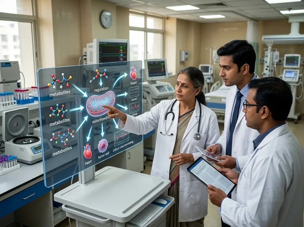
A groundbreaking review highlights mitochondrial quality control as a pivotal gating layer between metabolite signaling and regulated cell death, offering transformative therapeutic avenues across cardiology, oncology, and critical care.
Today
