.webp&w=384&q=75)

Loading, please wait...
Loading, please wait...

The surgical management of conductive hearing loss presents intricate microsurgical challenges, making the development of accurate 3D-printed human ossicles an important frontier in otolaryngology. Additive manufacturing offers unprecedented opportunities to reproduce delicate middle ear structures for surgical simulation, resident dissection training, and personalized prosthesis design. However, replicating submillimeter ossicular anatomy requires meticulous technological integration across imaging, computer-aided design, and additive manufacturing workflows.
Recent systematic reviews demonstrate surging clinical and academic interest in applying additive manufacturing to otology. Specifically, researchers have evaluated dozens of middle ear fabrication models worldwide. Most published studies reproduce ossicles embedded within healthy temporal bones, whereas models depicting pathological anatomy or isolated ossicular chains remain scarce. This trend reflects the technical ease of segmenting normal radiological scans compared to diseased ears with middle ear cholesteatoma or chronic erosive changes. Furthermore, otologists utilize these physical models primarily to rehearse delicate middle ear interventions, investigate acoustic biomechanics, and test custom reconstructive prostheses. Recreating genuine ossicular geometry provides realistic tactile feedback that screen-based virtual simulations cannot deliver. Therefore, high-fidelity physical models bridge the gap between complex theoretical anatomy and intricate microsurgery. However, existing literature frequently lacks standardized validation protocols, limiting direct comparisons across studies. Otolaryngology educators increasingly emphasize the need for uniform benchmarks before embedding these models routinely into residency curricula.
Fabricating anatomically faithful ossicular constructs fundamentally depends on the spatial resolution of the source radiological acquisition. Standard multislice computed tomography often fails to capture the fragile crura and footplate of the stapes accurately. Consequently, investigators increasingly utilize high-resolution cone-beam computed tomography and micro-computed tomography to acquire pristine volumetric datasets. These modalities reliably delineate the fine boundaries of the malleus head, the incus body, and the delicate stapes superstructure. In addition, the subsequent image segmentation phase introduces significant technical variability that directly alters final print dimensions. Common artifacts, including partial volume averaging and operator thresholding subjectivity, frequently expand or contract delicate ossicular contours during digital rendering. Therefore, otologists and imaging specialists must implement semi-automated, standardized thresholding algorithms to minimize human error. Without rigorous radiological post-processing controls, even industrial printers reproduce distorted models that mislead trainees during delicate dissection simulations. Precise segmentation protocols therefore serve as the foundational cornerstone for successful downstream additive manufacturing.
The choice of additive manufacturing platform directly determines the dimensional accuracy and surface resolution of 3D-printed middle ear models. Historically, material extrusion techniques such as fused deposition modeling lacked the fine resolution necessary to replicate tiny ossicular features. Today, photopolymerization technologies dominate the field, particularly stereolithography and material jetting, commonly known as PolyJet printing. These liquid resin platforms achieve ultra-thin layer heights, preserving critical landmarks such as the lenticular process and the stapedial arch. Moreover, PolyJet systems enable simultaneous multi-material deposition, allowing developers to fabricate flexible simulated ligaments alongside rigid bony structures. Clinicians can thus interact with realistic, multi-density temporal bone specimens that mirror living surgical fields under the operating microscope. Nevertheless, photopolymer resins require post-print chemical washing and ultraviolet light curing. These post-curing steps occasionally induce material shrinkage or structural warpage, which alters final prosthesis dimensions. Hence, engineering teams must calibrate resin exposure parameters carefully to maintain submillimeter geometric tolerances during manufacturing.
Across systematic evaluations of middle ear models, the stapes consistently represents the most difficult ossicle to replicate successfully. The malleus and incus possess substantial bony volume, facilitating reliable segmentation and physical printing in most simulations. Conversely, the stapes features an exceptionally thin arch and an ultra-thin footplate measuring mere fractions of a millimeter. Standard printers often obliterate the delicate obturator foramen or fail to cure the fragile crura, producing incomplete models. Furthermore, functional hearing requires unimpeded mechanical mobility between the ossicles, which are connected by the incudomalleolar and incudostapedial synovial joints. Monomaterial rigid prints fuse these articulations together, creating a static structure that fails to mimic genuine acoustic conduction. To overcome this limitation, innovative investigators incorporate elastomeric resins or construct micro-articulated joint spaces to restore lifelike chain mobility. Achieving realistic movement enables trainees to practice authentic ossicular palpation and trial partial or total replacement prostheses under realistic conditions. Consequently, dynamic multi-material printing represents a crucial advancement in otologic simulation.
Otolaryngology residency training faces growing constraints regarding cadaveric temporal bone access, procurement costs, and biohazard compliance. In this context, 3D-printed anatomical models provide an accessible, ethical, and fully repeatable alternative for microsurgical education. Novice residents use these synthetic replicas to practice surgical navigation, mastoidectomy, and middle ear exploration without risking patient safety. Furthermore, magnified models allow trainees to master delicate micro-instrumentation skills before attempting life-sized ossicular dissections. In reconstructive surgery, patient-specific 3D printing holds tremendous potential for custom ossicular replacement prostheses that fit irregular middle ear cavities perfectly. However, clinical translation requires overcoming current reporting deficiencies in published research. Investigators rarely publish detailed specifications regarding segmentation thresholds, printing tolerances, or objective surgical assessment metrics. Therefore, academic surgical societies must establish standardized consensus reporting guidelines for additive manufacturing in otology. Developing robust validation frameworks will accelerate regulatory clearance, refine surgical training curricula, and ultimately improve auditory outcomes for patients undergoing ossiculoplasty.
The stapes is the smallest bone in the human body, presenting an exceptionally delicate arch and a footplate measuring fractions of a millimeter. Standard imaging modalities and 3D printers struggle to resolve these microscopic dimensions without causing structural distortion or complete absence of the crura. Furthermore, the obturator foramen frequently clogs with unpolymerized resin during manufacturing, requiring specialized high-resolution micro-computed tomography and advanced stereolithography to achieve true anatomical fidelity.
Vat photopolymerization methods, particularly stereolithography and material jetting PolyJet systems, deliver the highest anatomical realism for middle ear replication. These technologies utilize liquid photopolymer resins that cure under targeted light beams, producing ultra-thin layers with micron-level resolution. Moreover, PolyJet platforms can combine multiple resins simultaneously, enabling developers to simulate flexible joint ligaments alongside rigid bony ossicles. This combination yields superior surface smoothness, dimensional precision, and authentic tactile handling for otologic simulation.
Currently, most 3D-printed ossicular models serve primarily educational, training, and biomechanical research purposes rather than routine human implantation. While biocompatible titanium and ceramic printing technologies show immense clinical promise for patient-specific reconstructive implants, regulatory clearance, long-term biocompatibility data, and commercial scalability remain active hurdles. Standard manufactured titanium prostheses continue to represent the clinical benchmark in ossiculoplasty until individualized additive manufacturing protocols undergo extensive prospective clinical trial validation.
Disclaimer: This content is for informational and educational purposes only and does not constitute formal medical advice, diagnosis, or treatment. Healthcare providers should exercise their independent clinical judgment and correlate these findings with specific clinical presentations and institutional protocols. Refer to the latest local and national guidelines for clinical practice.
References

Read summarized clinical updates, watch expert medical content, and earn CME certifications right from your smartphone.
Open your smartphone camera to scan and install.





A systematic review explores the current status of 3D-printed human ossicles, examining micro-CT imaging, stereolithography, PolyJet fabrication, and stapes replication challenges for surgical training and middle ear reconstruction.
Today
A multicity study highlights that over 80% of acute respiratory infections remain outside hospitals. Patients frequently choose community pharmacies and digital platforms. Integrating digital health data and retail pharmacy sales can fill critical surveillance gaps and improve early outbreak detection.
Today

Scandinavian register evidence reveals that neonatal birth length strongly predicts adult health and mortality, whereas birth weight dictates cognitive capacity. Symmetric growth restriction causes far more severe long-term deficits than asymmetric restriction, underscoring the need for comprehensive anthropometry.
Today
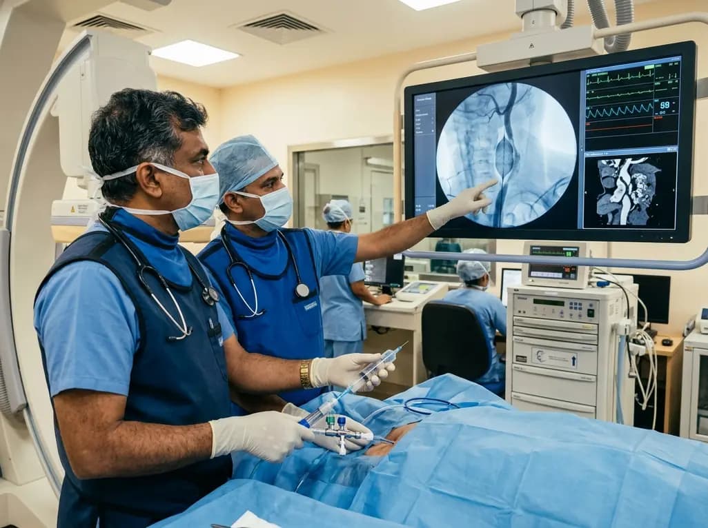
Discover how rescue cutting balloon angioplasty successfully modifies heavily calcified carotid stenosis, enabling stent delivery without baroreceptor instability and ensuring favorable long-term neurologic outcomes.
Today
A breakthrough study reveals that digoxin alleviates osteoarthritis by targeting LSP1 in synovial macrophages, preventing harmful extracellular trap formation and restoring SOD1 activity. This dual-action mechanism protects joint cartilage and offers a promising repurposing strategy for disease modification.
Today
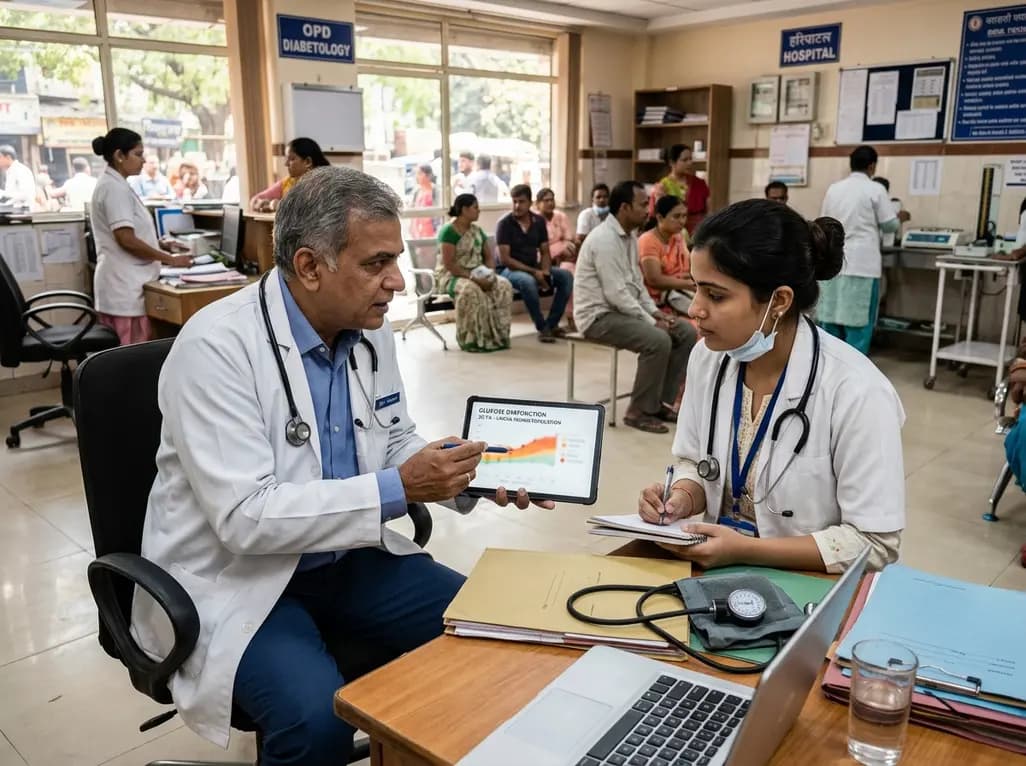
Data from the 2026 Workforce Health Index reveals that 38.7% of urban employees and dependents exhibit abnormal glycemic control. Emerging at increasingly younger ages, this metabolic shift intersects with severe vitamin deficiencies, highlighting the urgent need for early workplace screenings and targeted clinical care.
Today
