.webp&w=384&q=75)

Loading, please wait...
Loading, please wait...
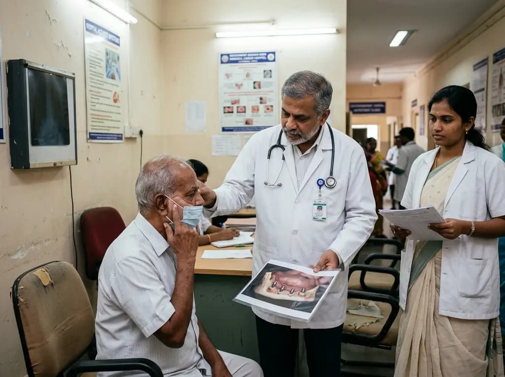
Surgical extirpation of head and neck malignancies frequently results in extensive tissue loss, compromising facial appearance, speech, and masticatory function. While autologous microvascular reconstruction represents the standard approach for many defects, complex auricular, orbital, and midfacial deficits often necessitate prosthetic reconstruction. Clinicians have long relied on osseointegrated hardware to secure retention. However, newly published data challenge historic assumptions regarding long-term craniofacial implant survival in oncologic cohorts, demonstrating that real-world outcomes diverge sharply from non-cancer populations.
Historically, maxillofacial prosthodontists and surgical teams referenced landmark literature indicating implant retention rates above 85% to 90%. However, investigators derived those historic figures primarily from heterogeneous patient cohorts, including individuals with congenital microtia or post-traumatic tissue loss. Conversely, oncologic patients present a vastly different local and systemic physiology. A retrospective study tracking 184 bone-anchored implants in 51 oncologic patients across a UK center revealed sobering results. The study recorded cumulative retention rates of 94% at one year, dropping to 60% at three years, and plunging to 46% at five years.
Consequently, the actual five-year craniofacial implant survival in this cancer population proved to be approximately half of traditional expectations. Moreover, the median survival stood at only 4.1 years. These figures indicate that surgical teams cannot extrapolate general craniofacial data to post-ablative cancer patients without substantially misleading clinical prognoses.
Radiation therapy remains an indispensable modality in curative head and neck oncology, yet it introduces profound structural damage to native bone. In the UK cohort, 77% of patients received adjuvant or definitive radiation, with a median delivered dose of 57 Gy to the implant bed. High-dose ionizing radiation induces progressive hypovascularity, hypocellularity, and tissue hypoxia, establishing severe microvascular obliterative endarteritis.
Furthermore, radiation hampers normal bone remodeling cycles by impairing osteoblast activity and triggering persistent fibroatrophy. Univariate models confirmed that radiation exposure serves as a prominent predictor of implant loss. Although multivariable testing displayed a statistical trend toward failure rather than an absolute isolated hazard, the biological reality of radiation-induced bone damage remains indisputable. Over time, tissue friability and chronic ischemia undermine the bone-to-implant contact interface, creating vulnerability to mechanical loosening and secondary infections.
In addition to radiation sequelae, systemic metabolic dysregulation critically threatens hardware longevity. On multivariable regression analysis, pre-existing diabetes emerged as an exceptionally powerful independent predictor of implant loss, carrying a fourfold increased hazard (HR 4.0, 95% CI 1.5–10.7). Even small cohorts of diabetic individuals experience disproportionately accelerated failure curves following surgical reconstruction.
Hyperglycemia impairs neutrophil chemotaxis, blunts bactericidal clearance, and fosters systemic microangiopathy that compounds radiation damage. Older age also correlated with premature failure in univariate analysis, reflecting attenuated osteogenic regenerative reserves. Therefore, oncologic teams operating within regions with high metabolic disease burdens must rigorously screen and optimize glycemic indices prior to inserting retentive fixtures, recognizing that diabetic microvascular changes markedly diminish the bone bed's structural tolerance.
Standard dental implant therapy in healthy bone typically reaches a survival plateau after initial osseointegration and loading, exhibiting minimal loss beyond the first 12 to 24 months. In striking contrast, the hazard analysis for bone-anchored oncologic fixtures demonstrated persistent, linear attrition without establishing an asymptote. Patients continued to lose osseointegrated anchors throughout the entire 20-year follow-up window.
This persistent downward trajectory reflects an unremitting biological toll. The combination of chronic percutaneous interface stress, ongoing radiation fibroatrophy, soft tissue thinning, and progressive bone resorption creates continuous instability. Consequently, successful primary healing provides zero guarantee against failure years later. Prosthetists and maxillofacial surgeons must anticipate late mechanical and biological breakdowns as an expected manifestation of irradiated osseous tissue, rather than treating late failure as an unexpected anomaly.
These findings necessitate immediate updates to reconstructive and prosthodontic protocols. Because late failure rates are pervasive, surgical teams must adopt a preemptive philosophy by placing supernumerary implants during primary or staged reconstruction. By placing redundant, strategically spaced bone fixtures, clinicians build functional redundancy into the prosthetic framework. If a single supporting fixture fails, the prosthesis remains anchored without requiring extensive revision surgery in compromised tissue.
Additionally, patient counseling must convey realistic long-term expectations rather than overpromising permanent hardware stability. Surveillance cannot cease once the surgical wound matures and the prosthesis is fitted. Instead, oncologic teams must mandate lifelong follow-up intervals to debride peri-implant tissues, monitor hygiene, manage cutaneous cuff inflammation, and adjust mechanical load distribution before localized bone resorption destabilizes the entire reconstructive framework.
Unlike congenital or trauma patients, oncologic patients frequently receive high-dose ionizing radiation and undergo extensive vascular alteration. Radiation creates enduring bone hypoxia, hypocellularity, and progressive fibroatrophy, which steadily erodes osseointegration over time. This ongoing biological strain prevents bone stabilization, causing failure rates to climb steeply between years three and five.
Diabetes induces systemic microangiopathy that severely diminishes microvascular perfusion, compounding the ischemia caused by surgical dissection and radiotherapy. Furthermore, hyperglycemia impairs neutrophil function and blunts collagen synthesis, which weakens peri-implant soft-tissue seals. These metabolic deficits facilitate opportunistic bacterial colonization and bone resorption, elevating the hazard of hardware loss fourfold.
Surgical teams increasingly install supernumerary or redundant implants during initial reconstruction rather than minimal retentive units. Providing extra fixtures ensures that if individual anchors fail over time, the remaining implants can still stabilize the facial prosthesis. This preventive design avoids repeated complex operations in chronically irradiated bone.
Disclaimer: This content is for informational and educational purposes only and does not constitute formal medical or dental advice. Clinical decisions for head and neck oncologic reconstruction should be made by qualified surgical and prosthetic specialists following comprehensive multidisciplinary evaluation. Refer to the latest local and national guidelines for clinical practice.
References

Read summarized clinical updates, watch expert medical content, and earn CME certifications right from your smartphone.
Open your smartphone camera to scan and install.





A long-term study indicates that bone-anchored craniofacial implant survival in oncologic patients drops to 46% at five years. Learn how radiation therapy and diabetes drive continual attrition, demanding proactive prosthetic planning and supernumerary placement to optimize facial rehabilitation.
4 weeks back
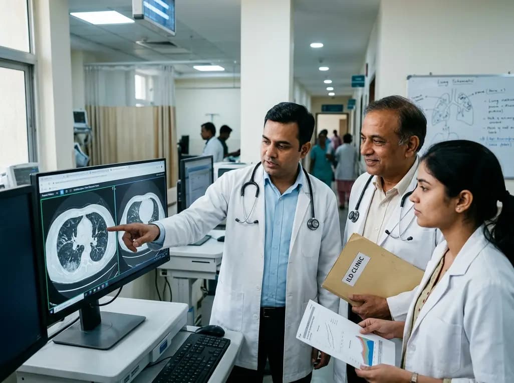
A nationwide English cohort study shows that interstitial lung diseases are no longer rare, with incidence jumping 30% and 200,000 active cases. This analysis highlights clinical implications for diagnosis, multi-disciplinary management, and global health priorities across respiratory and internal medicine.
Today

A nationwide South Korean study shows underreporting of smoking among women dropped from 57.4% to 36.4% between 2008 and 2021. However, over one-third of female smokers still conceal their habit, highlighting the critical role of cotinine verification in clinical risk assessment.
Today

PepsiCo and Monster Beverage have challenged the FSSAI ban on the energy drink label in Indian courts. While companies highlight supply disruptions, regulatory actions spotlight severe cardiovascular and metabolic risks associated with excessive caffeine, sugar, and taurine consumption, especially in adolescents.
Today

A new study evaluates the rheological stability of decellularized peripheral nerve matrix hydrogels under varied storage conditions. Findings show -80°C storage and freeze-drying preserve gelation kinetics and mechanical stiffness, offering key insights for shipping and translational clinical applications.
Today

A Bayesian multilevel meta-analysis reveals that aerobic training combined with moderate carbohydrate restriction modestly lowers HbA1c in type 2 diabetes. However, sparse data and very low certainty leave incremental benefits over exercise or diet alone unproven, highlighting the need for individualized care.
Yesterday
