.webp&w=384&q=75)

Loading, please wait...
Loading, please wait...
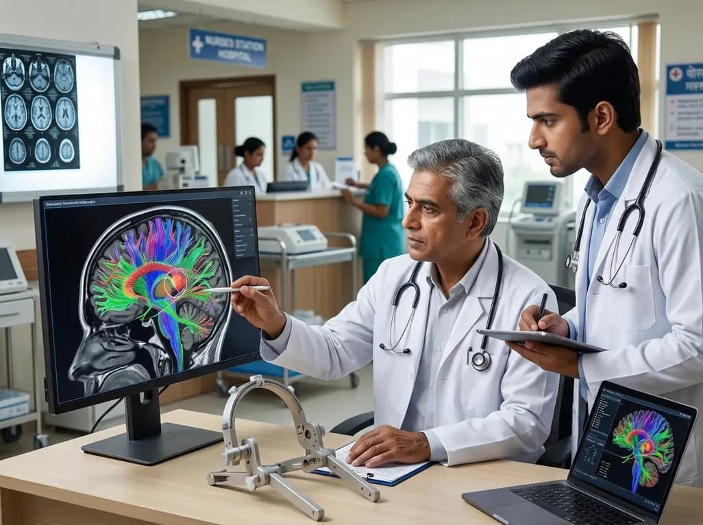
Essential tremor and tremor-dominant Parkinson disease cause profound disability among patients worldwide. While pharmacological therapy serves as the initial treatment line, many individuals eventually develop medical refractoriness. In such scenarios, clinicians frequently turn to surgical interventions to restore functional independence. Over recent decades, stereotactic radiosurgery thalamotomy has emerged as a compelling, non-invasive therapeutic modality for patients who are poor candidates for open neurosurgical procedures. By delivering highly focused ionizing radiation to specific thalamic targets, radiosurgery creates a precise, circumscribed lesion that disrupts aberrant oscillatory tremor circuits without requiring a craniotomy or hardware implantation.
Despite its proven efficacy and favorable safety profile, clinical outcomes following radiosurgical thalamotomy historically exhibit variable success rates. Consequently, clinicians have sought reliable anatomical and functional targeting biomarkers to standardize response rates. Standard stereotactic protocols often depend on indirect atlas-based coordinates. However, atlas targeting fails to account for inter-individual neuroanatomical variability and structural network differences. To overcome these limitations, advanced neuroimaging techniques now allow functional and structural connectome mapping. This paradigm shift offers an unprecedented opportunity to personalize treatment trajectories and improve tremor suppression outcomes.
Functional neurosurgery has increasingly embraced network-based medicine. Deep brain stimulation (DBS) protocols routinely utilize structural connectivity maps to optimize lead placement and stimulation parameters. In contrast, stereotactic radiosurgery thalamotomy has traditionally lagged in adopting these patient-specific connectomic methodologies. Most radiosurgical platforms rely primarily on structural magnetic resonance imaging (MRI) aligned with stereotactic coordinates referencing the anterior commissure-posterior commissure (AC-PC) line. Because the ventral intermediate nucleus (VIM) lacks distinct contrast boundaries on conventional T1- or T2-weighted MRI sequences, stereotactic placement remains susceptible to spatial imprecision.
Therefore, integrating probabilistic tractography into radiosurgical planning bridges a vital clinical gap. By mapping the axonal pathways traversing the thalamocortical network, tractography identifies the unique structural fingerprints of individual patients. Structural connectivity accurately delineates the sub-regions of the motor thalamus that communicate directly with key sensorimotor cortical hubs. Consequently, radiosurgical planning shifts away from static population averages toward dynamic, patient-tailored neural circuits. This connectomic strategy ensures that radiation delivers maximum biological effect to pathologically synchronized networks while sparing adjacent critical sensory and internal capsule pathways.
A pivotal retrospective study evaluated 27 patients enrolled in a prospective trial utilizing frameless virtual-cone radiosurgery on a linear accelerator. The cohort included individuals suffering from medically refractory essential tremor or tremor-dominant Parkinson disease. Investigators assessed the therapeutic response using percentage improvements in the contralateral Fahn-Tolosa-Marin Tremor Rating Scale (FTMTRS) scores over an average follow-up period of 17.9 months. The scientific team aimed to identify whether individual thalamocortical connectivity patterns directly correlated with optimal tremor suppression.
To evaluate these network connections, researchers performed advanced probabilistic tractography on high-resolution diffusion MRI scans. Specifically, they measured connectivity profiles from every thalamic voxel to three distinct cortical zones: the primary motor cortex (M1), the primary sensory cortex (S1), and the supplementary motor area/premotor cortex (SMA/PMC). Furthermore, the investigators conducted group-level comparisons to identify the statistical "sweet spot" for maximum contralateral tremor relief. Finally, multiple regression models evaluated the spatial distance between individual radiation lesion centers and the voxels displaying maximal connectivity to these defined cortical targets.
The study yielded remarkable insights into the neurobiological determinants of radiosurgical success. At the group level, the therapeutic sweet spot for maximum contralateral tremor relief aligned directly within the thalamic zone displaying peak structural connectivity to the primary motor cortex (M1). Furthermore, single-subject analyses revealed a statistically significant relationship between spatial targeting accuracy and clinical improvement. Specifically, smaller distances along the anterior-posterior axis between the radiosurgical lesion centerpoint and the voxel maximally connected to M1 correlated strongly with superior tremor relief.
In contrast, spatial distances relative to voxels maximally connected to the primary sensory cortex (S1) or SMA/PMC did not demonstrate a significant correlation with tremor suppression. These critical findings establish that effective stereotactic radiosurgery thalamotomy relies predominantly on interrupting specific M1-projecting thalamocortical tracts. Because individual variations in tract anatomy alter the exact coordinates of this optimal target, patient-specific tractography provides clinicians with an indispensable targeting biomarker. This precision minimizes off-target radiation delivery, thereby reducing risks of sensory paresthesias or motor deficits.
Incorporating connectomic workflows into routine neurosurgical and radiation oncology practices represents a vital evolutionary step. Currently, functional neurosurgeons often balance invasive DBS, magnetic resonance-guided focused ultrasound (MRgFUS), and radiosurgery. For patients with high surgical risk, advanced age, or anticoagulant dependencies, stereotactic radiosurgery remains an exceptional treatment choice. However, the delayed onset of radiation-induced biological effects makes accurate initial targeting imperative, as clinicians cannot adjust parameters intraoperatively in real time.
By leveraging patient-specific M1 tractography before linear accelerator or Gamma Knife delivery, multidisciplinary teams can customize beam geometries to match the patient's individual functional anatomy. Consequently, this refined method optimizes therapeutic response while preserving healthy brain architecture. Furthermore, standardizing tractography acquisition protocols across institutions will facilitate broader clinical adoption. Training radiation oncologists, functional neurosurgeons, and medical physicists in connectomic segmentation ensures that complex diffusion metrics translate smoothly into everyday stereotactic workflows.
As neuroimaging techniques evolve, the fusion of structural tractography with functional resting-state connectomics holds substantial promise for movement disorder management. High-field MRI systems and advanced diffusion algorithms will further refine the spatial resolution of thalamic sub-nuclei. Additionally, artificial intelligence algorithms may soon automate the delineation of individualized sweet spots, streamlining the entire radiosurgical planning process. Such automation will minimize inter-observer variability and expand access to personalized radiosurgery across diverse healthcare institutions.
Moreover, expanding connectomic research into bilateral tremor interventions, dystonia, and refractory epilepsy could transform functional radiosurgery. Multi-center prospective clinical trials will be essential to validate these findings across larger patient populations and diverse radiosurgical delivery platforms. By transitioning from rigid anatomical landmarks to patient-specific neural network targeting, functional radiosurgery is entering an era of true precision medicine. Ultimately, these technological innovations ensure that patients receive safer, more effective, and enduring relief from debilitating movement disorders.
Connectomic guidance enables clinicians to target patient-specific functional brain networks rather than relying on generic anatomical coordinates. This personalized approach identifies the precise thalamic region connected to the primary motor cortex, maximizing tremor reduction while significantly reducing radiation exposure to adjacent sensory and motor pathways.
Radiosurgical thalamotomy is a completely non-invasive, incisionless outpatient procedure that does not require hardware implantation or repeat programming. In contrast, deep brain stimulation requires surgical lead placement and battery maintenance, though it offers immediate symptom relief and reversible, adjustable stimulation parameters for medically eligible patients.
Unlike invasive surgical procedures that provide immediate tremor suppression, stereotactic radiosurgery relies on gradual biological tissue modulation. Consequently, patients typically observe progressive tremor reduction over several months following treatment, with peak clinical improvements generally emerging between six and twelve months post-procedure.
Disclaimer: This content is for informational and educational purposes only and is not intended to serve as medical advice, diagnosis, or treatment. Healthcare professionals should exercise their independent clinical judgment. Refer to the latest local and national guidelines for clinical practice.
References

Read summarized clinical updates, watch expert medical content, and earn CME certifications right from your smartphone.
Open your smartphone camera to scan and install.





A groundbreaking study highlights how connectomic-guided stereotactic radiosurgery thalamotomy improves tremor control by utilizing patient-specific structural connectivity to the primary motor cortex, paving the way for personalized non-invasive neurosurgery.
4 weeks back

Chronic eosinophilic pneumonia can present with atypical bronchoalveolar lavage findings and an organizing pneumonia pattern, necessitating surgical lung biopsy.
Today

Discover the crucial distinctions between mesonephric and mesonephric-like proliferations of the female genital tract. Learn the key histomorphologic, immunohistochemical, and molecular differences essential for accurate diagnosis and clinical management.
Today
Kikuchi-Fujimoto disease rarely presents with refractory shock and sinus bradycardia in children. This case report highlights life-threatening hemodynamic instability requiring intensive care, preserved cardiac contractility, and the critical diagnostic role of early excisional lymph node biopsy.
Today

A narrative review investigates whether the articularis genus muscle functions as an independent anatomical entity or blends with the vastus intermedius. We evaluate its morphology, role in retracting the suprapatellar bursa, and direct clinical significance in anterior knee pain and arthroplasty.
Today

A Bayesian multilevel meta-analysis reveals that aerobic training combined with moderate carbohydrate restriction modestly lowers HbA1c in type 2 diabetes. However, sparse data and very low certainty leave incremental benefits over exercise or diet alone unproven, highlighting the need for individualized care.
Today
