.webp&w=384&q=75)

Loading, please wait...
Loading, please wait...
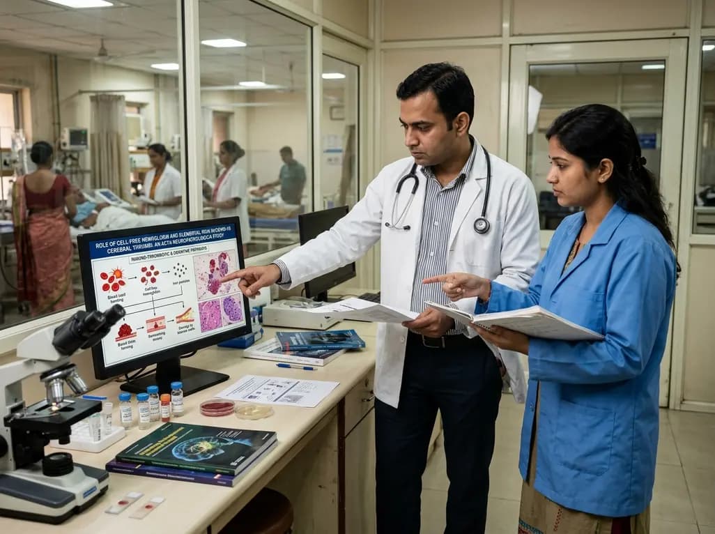
Ischemic stroke remains one of the most devastating complications observed during acute SARS-CoV-2 infection. While clinicians quickly recognized the hypercoagulable state of COVID-19 early in the pandemic, the exact molecular architecture within occlusive vascular clots remained elusive. Emerging translational evidence now demonstrates that cell-free hemoglobin and elemental iron play a pivotal role in driving this persistent prothrombotic condition. By examining retrieved clots from acute ischemic stroke patients, investigators have uncovered unique biophysical and biochemical signatures that distinguish viral immunothrombosis from typical thromboembolic events. Consequently, understanding these intricate vascular pathways provides clinicians with essential insights into targeted therapeutics and post-viral cardiovascular management.
Cerebral clots retrieved via mechanical thrombectomy during the pre-Omicron pandemic phase exhibit distinct morphological properties. Specifically, transmission electron microscopy reveals a markedly disorganized fibrillar network throughout COVID-19 thrombi compared to non-infected historical controls. Standard histopathological evaluations using Masson's trichrome staining further corroborate these findings by demonstrating an irregular, less densely packed extracellular matrix. Furthermore, this abnormal structural porosity alters clot stability and biomechanical resistance against endogenous fibrinolytic enzymes. In addition, immunohistochemical profiling indicates an unusual distribution of immune cells within the clot environment. Although neutrophil extracellular traps (NETs) and erythrocyte counts appear relatively comparable across cohorts, COVID-19 thrombi show a significant trend toward elevated macrophage infiltration. Specifically, these samples display a diffuse CD68-positive cellular pattern accompanied by a marked reduction in total platelet content. Therefore, these structural modifications reflect an atypical thrombo-inflammatory process rather than standard platelet-driven arterial aggregation.
Quantitative proteomics using liquid chromatography-tandem mass spectrometry provides critical molecular clarity regarding clot composition. Among 720 shared proteins analyzed across samples, researchers identified 48 differentially expressed proteins that define the unique COVID-19 thrombotic signature. Most notably, the data demonstrate a striking upregulation of multiple hemoglobin subunits, including alpha, beta, gamma, and delta chains. Simultaneously, essential clearance molecules such as haptoglobin and biliverdin reductase show substantial enrichment within the retrieved thrombi. Redox-regulating proteins also increase significantly, highlighting severe localized oxidative stress within the cerebral vasculature. In contrast, standard platelet structural and activation proteins are noticeably downregulated in COVID-19 clots. Crucially, glycophorin A immunostaining and structural erythrocyte marker analyses show no corresponding increase in intact red blood cells. Consequently, these findings prove that the elevated cell-free hemoglobin exists outside intact erythrocytes, accumulating directly within the extracellular matrix of the cerebral clot.
To quantify the inorganic elements within the retrieved neurovascular occlusions, investigators utilized total reflection X-ray fluorescence elemental profiling. This highly sensitive analytical technique confirmed significantly elevated concentrations of elemental iron within COVID-19-associated thrombi. When red blood cells undergo intravascular hemolysis or microvascular shear destruction, released heme molecules rapidly dissociate and release free ferrous and ferric ions. Consequently, unchelated iron catalyzes Fenton-type chemical reactions, driving the aggressive generation of toxic hydroxyl free radicals. Furthermore, this intense oxidative surge directly oxidizes surrounding fibrinogen molecules, causing them to polymerize into dense, fibrinolytic-resistant paracrystalline networks. Thus, the co-enrichment of cell-free iron and oxidized heme establishes a self-amplifying cascade of vascular injury. Moreover, this severe local oxidative environment exhausts endogenous scavengers, including local haptoglobin and hemopexin reserves. As a result, the cerebral microenvironment suffers extensive endothelial dysfunction, perpetuating thrombosis and worsening secondary ischemic neuronal damage.
Beyond hemolysis-driven pathways, proteomic investigations demonstrate a robust humoral and immune-mediated signature unique to viral-associated clots. Specifically, acute-phase reactants, classical complement components, and diverse immunoglobulin subclasses appear exclusively within COVID-19 thrombus samples. This distinct molecular profile illustrates how the systemic cytokine storm converges with local intravascular coagulation cascades. When SARS-CoV-2 triggers endothelial activation, circulating antibodies and immune complexes deposit within the damaged vessel wall. Subsequently, classical complement pathway activation amplifies endothelial detachment, exposing highly thrombogenic subendothelial basement membranes. Furthermore, CD68-positive macrophages recruited to these sites actively interact with free heme species, releasing additional pro-inflammatory cytokines and tissue factor. Therefore, the physical cerebral clot serves not merely as a mechanical vascular obstruction, but as an active, localized crucible of persistent immunothrombosis and chronic inflammation.
These breakthrough pathological findings carry profound clinical implications for neurologists, critical care specialists, and interventionists managing post-viral vascular syndromes. Standard antiplatelet agents such as aspirin and clopidogrel primarily target platelet activation and thromboxane synthesis. However, because COVID-19 thrombi display reduced platelet density alongside heavy cell-free heme and iron accumulation, standard antiplatelet therapy alone may prove suboptimal. Instead, therapeutic strategies could expand to incorporate targeted iron chelators, haptoglobin replacement therapy, or specialized free-radical scavengers to mitigate ongoing oxidative vascular injury. Furthermore, recognizing that atypical clot architecture alters mechanical properties may help neuro-interventional teams refine thrombectomy techniques and device selection. Additionally, clinicians evaluating high-risk infected patients should maintain vigilance for subclinical intravascular hemolysis and elevated systemic inflammatory markers. Consequently, neutralizing free heme toxicity and blunting complement-mediated vessel injury could emerge as crucial adjuvant strategies to prevent recurrent stroke.
While these analytical observations provide pivotal mechanistic clarity, they also open vital avenues for prospective multi-center validation. Future clinical investigations must evaluate whether circulating biomarkers of hemolysis, such as plasma cell-free hemoglobin and serum ferritin, directly correlate with stroke severity and recanalization success rates. Moreover, researchers should examine whether similar iron-driven immunothrombotic mechanisms occur in other major viral infections or post-acute sequelae. Investigating novel pharmacological agents that neutralize extracellular hemoglobin toxicity without impairing normal systemic hemostasis represents a high-priority translational goal. In addition, advanced neuroimaging modalities could eventually detect iron-rich, lytic-resistant thrombi non-invasively prior to mechanical intervention. Ultimately, bridging molecular proteomic discovery with bedside acute stroke care will empower clinicians to deliver personalized, biologically tailored therapies for complex infectious and inflammatory vascular disorders.
Cell-free hemoglobin releases unchelated elemental iron and reactive heme molecules directly into the cerebral vasculature. Consequently, this induces intense Fenton-driven oxidative stress, leading to endothelial damage, oxidative modification of fibrinogen, and the formation of dense, lysis-resistant clots that obstruct major cerebral arteries during severe viral infection.
Unlike standard arterial thrombi driven by primary platelet aggregation, COVID-19-associated clots stem predominantly from an immunothrombotic cascade. Massive immune-inflammatory activation, complement deposition, macrophage infiltration, and severe oxidative stress generate an irregular extracellular matrix composed mainly of cell-free hemoglobin, fibrin, and inflammatory proteins rather than densely packed platelet aggregates.
Identifying cell-free hemoglobin and elemental iron in thrombi suggests that adjuvant therapies, such as iron chelators, haptoglobin replenishment, and antioxidant scavengers, could protect the neurovascular endothelium. Combining these novel agents with standard anticoagulation or thrombectomy may reduce oxidative reperfusion injury and improve long-term functional recovery after ischemic stroke.
Disclaimer: This content is for informational and educational purposes only and should not be construed as medical advice. Clinical decisions must always be made by qualified healthcare professionals based on individual patient assessment. Refer to the latest local and national guidelines for clinical practice.
References
Payá M et al. High levels of cell-free hemoglobin and iron in cerebral thrombi of pre-Omicron COVID-19 stroke patients: novel drivers of SARS-CoV-2-induced prothrombotic state. Acta Neuropathol. 2026 Aug 29. doi: 10.1007/s00401-026-03074-7. PMID: 42667425.
Al-Samkari H, Karp Leaf RS, Dzik WH, et al. COVID-19 and coagulation: bleeding and thrombotic manifestations of SARS-CoV-2 infection. Blood. 2020;136(4):489-500. doi:10.1182/blood.2020006520.
Buehler PW, D'Agnillo F, Schaer DJ. Hemoglobin-driven pathophysiology is an emerging target in clinical medicine. Trends Mol Med. 2020;26(4):373-385. doi:10.1016/j.molmed.2019.11.006.

Read summarized clinical updates, watch expert medical content, and earn CME certifications right from your smartphone.
Open your smartphone camera to scan and install.





A new study in Acta Neuropathologica reveals that cerebral thrombi from COVID-19 stroke patients contain high levels of cell-free hemoglobin and elemental iron. These findings expose a distinct immuno-thrombotic and oxidative mechanism, offering novel targets for vascular protection and stroke therapeutics.
4 weeks back

A recent clinical case series highlights the synthetic polylactic acid matrix as an innovative local therapy for refractory pyoderma gangrenosum. The bioresorbable scaffold demonstrated complete ulcer closure and favorable scar quality across chronic wounds, presenting a viable regenerative modality.
Today

A nationwide South Korean study shows underreporting of smoking among women dropped from 57.4% to 36.4% between 2008 and 2021. However, over one-third of female smokers still conceal their habit, highlighting the critical role of cotinine verification in clinical risk assessment.
Today
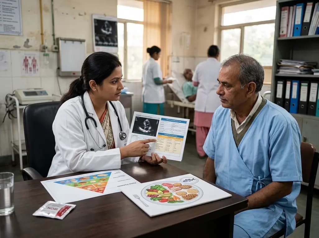
A scientific statement highlights the crucial role of nutrition in managing chronic heart failure. It emphasizes GLIM-based screening, individualized dietary patterns like the Mediterranean diet, appropriate protein and energy intake, avoiding unnecessary fluid restriction, and targeted iron supplementation.
Today

A new study evaluates the rheological stability of decellularized peripheral nerve matrix hydrogels under varied storage conditions. Findings show -80°C storage and freeze-drying preserve gelation kinetics and mechanical stiffness, offering key insights for shipping and translational clinical applications.
Today

A qualitative meta-synthesis highlights the complex interplay between gender dysphoria, minority stress, and eating disorders in transgender and gender-diverse individuals. Clinicians must recognize these distinct drivers to offer supportive, gender-affirming, and interdisciplinary eating disorder care.
Today
