

Loading, please wait...
Loading, please wait...

Recent neuroimaging investigations provide compelling clues regarding brain volume in IIH and its contribution to elevated intracranial pressure. Idiopathic intracranial hypertension primarily affects young women with elevated body mass index, presenting with severe headaches and papilledema. Historically, medical literature attributed this disorder entirely to cerebrospinal fluid dysregulation or impaired venous sinus outflow. However, newer volumetric magnetic resonance imaging analyses reveal a different dimension to this complex neurological syndrome. In particular, clinicians often struggle to explain why intracranial pressure spikes in the absence of mass lesions or overt hydrocephalus. Therefore, evaluating parenchymal changes offers critical mechanistic insights into the syndrome. Researchers at Duke University and the University of Rochester investigated whether parenchymal volume differences exist between affected individuals and healthy peers. Consequently, their observations challenge long-held assumptions regarding static brain parenchymal volume. Furthermore, the findings demonstrate that parenchymal expansion represents an active component of disease pathophysiology. As a result, clinicians must reconsider traditional pathophysiological paradigms to understand pressure gradients better. Ultimately, these quantitative discoveries pave the way toward refined diagnostic criteria and individualized treatment strategies for patients in routine clinical practice.
To examine parenchymal alterations, researchers retrospectively analyzed quantitative MRI scans from eighty-four patients with idiopathic intracranial hypertension alongside fifteen age-matched controls. Additionally, the cohort exhibited typical demographics, comprising eighty-seven percent female participants with a mean age of thirty years. The investigators applied automated volumetric segmentation algorithms to evaluate total brain volume, gray matter, and white matter. Furthermore, they normalized all measurements to total intracranial volume to prevent confounding from variations in cranial head size. The quantitative data revealed that patients with idiopathic intracranial hypertension had significantly higher total normalized brain volume than controls. In addition, affected individuals demonstrated expanded gray matter and white matter compartments compared to their healthy counterparts. Because obesity often influences metabolic parameters, the team controlled for age, sex, and body mass index during multiple linear regression. Consequently, parenchymal enlargement remained statistically significant even after adjusting for these demographic covariates. Thus, the expansion represents a true pathophysiological feature rather than a simple byproduct of patient body habitus. These objective neuroimaging metrics provide tangible evidence that parenchymal swelling plays an integral role in disease manifestation.
Although both gray and white matter compartments increased in volume, only gray matter demonstrated a direct association with cerebrospinal fluid opening pressure. Specifically, multiple linear regression analysis demonstrated a statistically significant positive correlation between expanding gray matter volume and opening pressure on lumbar puncture. In contrast, neither white matter volume nor total brain volume exhibited a statistically significant relationship with intracranial pressure. Therefore, cortical and deep gray structures appear uniquely sensitive to fluid dynamics within the cranium. Scientists hypothesize that diffuse intracellular astroglial swelling or microvascular congestion primarily affects the metabolic demand of cortical regions. Moreover, impaired glymphatic clearance through the brain parenchyma may cause extracellular interstitial fluid accumulation within gray matter. Because the rigid cranial vault limits volume expansion according to the Monro-Kellie doctrine, even modest parenchymal fluid retention raises intracranial pressure substantially. Consequently, gray matter expansion may directly provoke the symptomatic spikes in pressure that patients experience during disease progression. In addition, this selective volumetric shift provides clinicians with an objective anatomical explanation for fluctuating cerebrospinal fluid measurements during lumbar punctures.
Beyond opening pressure, the study established a compelling relationship between parenchymal volume and ophthalmic morbidity. Notably, patients presenting with severe optic disc swelling had significantly higher gray matter volume than patients presenting with mild papilledema. Because papilledema poses the greatest threat of irreversible vision loss in idiopathic intracranial hypertension, this correlation holds vital prognostic significance. Ophthalmologists routinely encounter patients whose visual fields deteriorate rapidly despite conventional medical management. Furthermore, higher gray matter volume likely amplifies transmission of intracranial pressure through the optic nerve sheath into the retrobulbar subarachnoid space. Consequently, axoplasmic flow stasis within retinal ganglion cells accelerates, driving severe optic disc edema and potential visual loss. In addition, the absence of white matter correlation suggests that parenchymal fluid shifts uniquely correlate with visual pathway strain. Therefore, neuroimaging volumetric markers could identify individuals at heightened risk for progressive visual compromise before severe visual field defects appear. Accordingly, incorporating parenchymal volume assessments into standard neuro-ophthalmic workups may help clinicians stratify ophthalmic risks more effectively.
These quantitative radiological insights introduce practical considerations for neurologists, ophthalmologists, and radiologists managing idiopathic intracranial hypertension. Traditionally, clinicians treat this condition primarily by decreasing cerebrospinal fluid production with carbonic anhydrase inhibitors like acetazolamide. However, medical teams should recognize that parenchymal fluid retention may resist standard fluid drainage therapies. Furthermore, this study underscores the necessity of high-resolution structural MRI scans during initial patient evaluation. While qualitative radiological reads often report normal brain parenchyma, subtle volumetric enlargement can escape visual inspection alone. Consequently, advanced quantitative volumetric post-processing tools could enhance diagnostic precision in borderline or atypical presentations. In addition, identifying high gray matter volume could prompt earlier, aggressive interventions to safeguard visual function. For example, neuro-ophthalmologists might recommend surgical decompression or venous sinus stenting more rapidly when high parenchymal volume coincides with refractory papilledema. Thus, evaluating the entire intracranial compartment helps clinicians move beyond simplistic fluid models toward holistic neurovascular management. Ultimately, personalized clinical care will depend on integrating parenchymal volumetric data with functional ophthalmic assessments.
According to the Monro-Kellie doctrine, the rigid skull accommodates fixed volumes of brain tissue, blood, and cerebrospinal fluid. When gray matter volume expands due to intracellular edema or microvascular congestion, intracranial compliance decreases sharply. Consequently, this parenchymal expansion directly elevates intracranial pressure and exacerbates characteristic compressive neurological symptoms.
Gray matter possesses high vascularity and metabolic activity, making it susceptible to interstitial fluid accumulation and glymphatic congestion. This expansion elevates cerebrospinal fluid opening pressure, transmitting mechanical stress directly into the optic nerve sheath. Consequently, heightened retrobulbar pressure disrupts axonal transport, aggravating optic disc swelling and causing severe papilledema.
Standard clinical MRI interpretation rarely detects subtle volumetric enlargement because radiologists assess images qualitatively for mass lesions or ventriculomegaly. Instead, identifying these alterations requires quantitative volumetric analysis using automated segmentation software normalized for head size. In the future, automated neuroimaging tools may integrate into routine workflows to assist clinical risk assessment.
Disclaimer: This content is for informational and educational purposes only... Refer to the latest local and national guidelines for clinical practice.
References
Schartz D et al. Increased brain volume in idiopathic intracranial hypertension: Association with intracranial pressure and papilledema severity. J Neurol Sci. 2025 Jun 15. doi: 10.1016/j.jns.2025.123501. PMID: 40239382.
Garza Hernandez S, Yordi S, Gratton SM. Diagnosis and Management of Idiopathic Intracranial Hypertension. EyeNet Magazine. 2025.
Mollan SP, Davies B, Silver NC, et al. New horizons in the diagnosis and management of idiopathic intracranial hypertension. Lancet Neurol. 2018;17(11):1021-1031.
Bender MT, Schartz D, Finkelstein A. A unifying disease model of idiopathic intracranial hypertension: a narrative review. Headache. 2026;66(4):990-995.

Read summarized clinical updates, watch expert medical content, and earn CME certifications right from your smartphone.
Open your smartphone camera to scan and install.





A recent volumetric MRI study reveals that patients with idiopathic intracranial hypertension have increased brain volume, with gray matter expansion directly correlating with opening pressure and papilledema severity, offering novel insights into disease pathophysiology.
Today

The Food Safety and Standards Authority of India has issued an urgent advisory against food business operators violating mandatory labelling standards for nutraceuticals and botanicals. Regulators prohibited therapeutic claims while mandating clear advisory warnings to prevent patient toxicity and medical misuse.
Today

Biotechnology researchers in India are developing an automated liquid biopsy platform to detect cervical malignancy from blood samples. Supported by the MAHA MedTech Mission, this initiative applies nanopore sequencing and piRNA biomarker detection to eliminate invasive speculum exams and expand rural screening.
Today
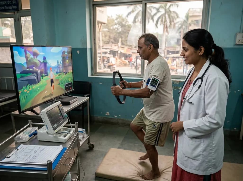
A clinical study evaluated Ring Fit Adventure in adults with chronic conditions, revealing that exergaming achieves moderate-to-vigorous exercise intensity while maintaining hemodynamic stability, high enjoyment, and zero adverse events.
Today

A retrospective cohort study reveals that selecting the touched vertebra as the lowest instrumented vertebra in thoracic adolescent idiopathic scoliosis yields a 20.8% distal add-on rate, driven by remnant motion segments and axial rotational discrepancies.
Today
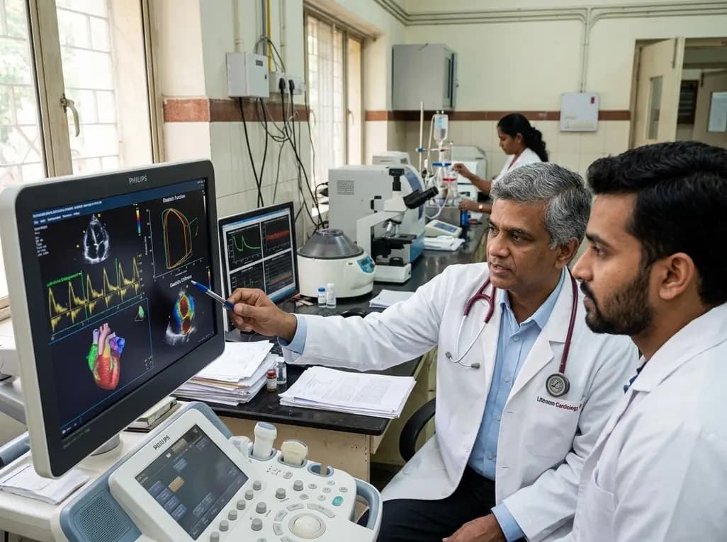
A study evaluates a cardiometabolic HFpEF model combining metabolic stress with chronic mineralocorticoid excess. The findings reveal profound diastolic stiffness, endothelial dysfunction, and pulmonary hypertension, illuminating key pathophysiological mechanisms for targeted cardiopulmonary therapies.
Today
