.webp&w=384&q=75)

Loading, please wait...
Loading, please wait...
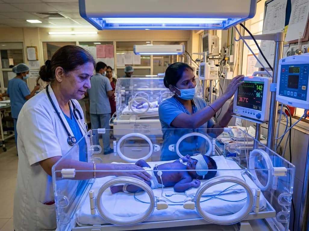
The neonatal period represents a critical window of vulnerability and rapid maturation for newborn infants. During this early developmental stage, the skin serves as an indispensable physical shield and an active immunological interface. Clinicians worldwide routinely utilize blue light phototherapy to treat neonatal hyperbilirubinemia, effectively converting toxic unconjugated bilirubin into excretable photoisomers. Consequently, this intervention has prevented bilirubin encephalopathy in millions of infants over several decades. However, emerging dermatological research indicates that phototherapeutic light exposure is not merely an inert, superficial therapy. Instead, blue light phototherapy directly interacts with dynamic cutaneous structures, triggering biological alterations that extend beyond simple bilirubin clearance. Understanding these subtle cutaneous interactions is vital for modern neonatal care.
Blue light phototherapy utilizes narrow-spectrum visible light, generally between 430 and 490 nanometers, to penetrate the epidermis and superficial dermis. At these specific wavelengths, photons reach vascular beds within the skin to induce photoisomerization of bilirubin. Nevertheless, visible light absorption concurrently excites endogenous chromophores, including flavins, porphyrins, and cytochromes. As a result, this excitation generates low levels of reactive oxygen species within cutaneous tissue. Consequently, intracellular signaling pathways activate, causing cellular stress responses within developing keratinocytes and dermal fibroblasts. Furthermore, early neonatal skin lacks robust antioxidant defenses and mature stratum corneum architecture. Therefore, photo-oxidative stress may temporarily suppress local cellular proliferation and alter membrane fluidity. In addition, blue wavelengths can induce subtle modifications in cellular mitochondrial activity, thereby altering metabolic balance within delicate cutaneous layers. These cellular dynamics demonstrate that visible spectrum phototherapy behaves as a biologically active exposure that interacts directly with immature skin tissues.
The neonatal epidermal barrier undergoes substantial physiological refinement during the initial weeks of extrauterine life. Crucially, stratum corneum hydration and lipid organization dictate overall cutaneous barrier competency. When clinicians administer blue light phototherapy, thermal radiation and direct photon exposure can alter the delicate lipid matrix of the stratum corneum. Consequently, treated infants frequently exhibit measurable increases in transepidermal water loss during and immediately after therapeutic sessions. Furthermore, elevated evaporative loss compromises stratum corneum hydration, thereby precipitating transient cutaneous xerosis and superficial desquamation. In addition, alterations in epidermal surface pH occur, shifting the mantle away from its optimal mildly acidic state. Because enzymatic processes responsible for ceramide synthesis require an acidic environment, this pH disruption may temporarily delay lipid processing. Thus, clinicians must recognize that phototherapy-induced barrier perturbation increases cutaneous fragility, predisposing infants to subclinical microtrauma, environmental irritant penetration, and secondary inflammatory cascades during a sensitive developmental period.
Microbial colonization of the newborn skin begins immediately at delivery, establishing a diverse commensal ecosystem that educates early immune responses. However, blue light phototherapy exerts significant photochemical effects on colonizing bacterial species. Because blue wavelengths activate bacterial porphyrins to generate lethal reactive oxygen species, light exposure selectively inhibits specific microbial strains while sparing others. Consequently, phototherapy can induce acute shifts in cutaneous biodiversity, creating temporary dysbiosis in the neonatal cutaneous flora. For instance, populations of beneficial commensals like coagulase-negative Staphylococci and early Corynebacterium strains may decline, which facilitates transient overgrowth of opportunistic pathobionts. In addition, phototherapy-induced alterations in epidermal sebum production and surface moisture further disrupt the niche microenvironments required for stable microbial succession. Therefore, perturbation of pioneer microbial communities during this formative window may alter the developmental trajectory of the infant skin microbiome, potentially compromising its long-term defensive and immune-regulatory capabilities.
Beyond its physical protective role, neonatal skin operates as a complex neuro-immuno-endocrine organ that engages in continuous systemic communication. When blue light phototherapy irradiates developing cutaneous tissue, it modulates resident immune cells, including Langerhans cells, dermal dendritic cells, and mast cells. Specifically, light exposure can alter local cytokine profiles by promoting transient increases in pro-inflammatory mediators such as interleukin-1 beta, interleukin-8, and tumor necrosis factor-alpha. Concurrently, systemic circulation transports these cutaneous inflammatory signals to distant organ systems, including the developing gut, central nervous system, and systemic immune reservoirs. Furthermore, cutaneous nerve fibers express photoreceptive pathways that respond to visible wavelengths, potentially modulating neuroendocrine signaling cascades via the hypothalamic-pituitary-adrenal axis. Consequently, the cutaneous responses triggered by blue light phototherapy extend far beyond the skin envelope, demonstrating how local dermal interventions can modulate broad cross-system networks during early neonatal development.
Given the critical interplay between barrier function, microbial colonization, and immune maturation, early disruptions may yield lasting clinical consequences. In particular, pediatric investigators increasingly examine the relationship between blue light phototherapy exposure and the subsequent development of atopic dermatitis. When phototherapy disrupts stratum corneum integrity and shifts the skin microbiome toward dysbiosis, it creates an environment conducive to epicutaneous allergen sensitization. Consequently, altered antigen presentation can skew naive T helper cell differentiation toward a Th2-dominated phenotype, which characterizes atopic eczema. Moreover, epidemiological studies have explored potential associations between neonatal phototherapy and elevated risks of childhood asthma, allergic rhinitis, and allergic march progression. Although current evidence remains complex with potential confounding by underlying hyperbilirubinemia severity, the biological plausibility linking early barrier injury to immune-mediated disorders is compelling. Therefore, longitudinal cohorts are essential to establish definitive causal links between neonatal light exposure and chronic childhood inflammatory conditions.
Understanding the multi-organ biological effects of blue light phototherapy does not diminish its status as an indispensable, life-saving therapeutic modality. Rather, these insights encourage pediatricians and neonatologists to optimize phototherapy protocols while safeguarding developing skin. First, clinicians should adhere strictly to established bilirubin treatment thresholds to avoid unnecessary or prolonged light exposure. Second, healthcare teams must maintain rigorous supportive skincare practices, including diligent hydration monitoring and the judicious use of gentle, barrier-restoring emollients post-treatment. Third, continuous development of targeted LED delivery systems allows precise spectral dosing that minimizes superfluous thermal and radiative strain on delicate infant skin. In the future, comprehensive longitudinal studies must evaluate the long-term dermatologic outcomes of phototherapy cohorts into later childhood. By integrating deeper photobiological understanding into everyday neonatal workflows, clinicians can maximize therapeutic efficacy against severe hyperbilirubinemia while protecting the delicate cutaneous and systemic health of vulnerable newborns.
Blue light phototherapy selectively affects colonizing microorganisms through the photoexcitation of endogenous bacterial porphyrins, which generates antimicrobial reactive oxygen species. Consequently, this photochemical mechanism can reduce beneficial commensal bacterial diversity while permitting opportunistic pathobionts to proliferate. In addition, phototherapy alters epidermal surface pH and moisture levels, thereby disrupting the fragile microenvironmental niches required for normal, long-term microbial colonization during the critical neonatal window.
Emerging scientific evidence indicates that phototherapy-induced epidermal barrier disruption, elevated transepidermal water loss, and microbial dysbiosis may facilitate epicutaneous allergen penetration in susceptible infants. Consequently, this early barrier impairment can stimulate immune pathways that favor a Th2-skewed allergic response, potentially increasing the risk of developing atopic dermatitis. However, ongoing longitudinal studies are necessary to clarify causality and differentiate light-induced effects from underlying hyperbilirubinemia.
Clinicians should ensure strict adherence to evidence-based bilirubin treatment thresholds to prevent unnecessary or excessive phototherapeutic exposure in newborns. Furthermore, maintaining optimal systemic hydration, closely monitoring fluid balance, and evaluating skin integrity remain paramount during treatment. Following the completion of phototherapy sessions, healthcare providers can consider applying gentle, hypoallergenic, barrier-supportive emollients to restore stratum corneum hydration and support healthy epidermal barrier recovery.
Disclaimer: This content is for informational and educational purposes only and should not be considered medical advice. Always consult a qualified healthcare provider for specific clinical circumstances. Refer to the latest local and national guidelines for clinical practice.
References

Read summarized clinical updates, watch expert medical content, and earn CME certifications right from your smartphone.
Open your smartphone camera to scan and install.





Blue light phototherapy is essential for neonatal jaundice, yet emerging evidence reveals systemic impacts on epidermal barrier integrity, microbial colonization, and long-term immune-mediated conditions.
4 weeks back

Chronic eosinophilic pneumonia can present with atypical bronchoalveolar lavage findings and an organizing pneumonia pattern, necessitating surgical lung biopsy.
Today

Discover the crucial distinctions between mesonephric and mesonephric-like proliferations of the female genital tract. Learn the key histomorphologic, immunohistochemical, and molecular differences essential for accurate diagnosis and clinical management.
Today
Kikuchi-Fujimoto disease rarely presents with refractory shock and sinus bradycardia in children. This case report highlights life-threatening hemodynamic instability requiring intensive care, preserved cardiac contractility, and the critical diagnostic role of early excisional lymph node biopsy.
Today

A narrative review investigates whether the articularis genus muscle functions as an independent anatomical entity or blends with the vastus intermedius. We evaluate its morphology, role in retracting the suprapatellar bursa, and direct clinical significance in anterior knee pain and arthroplasty.
Today

A Bayesian multilevel meta-analysis reveals that aerobic training combined with moderate carbohydrate restriction modestly lowers HbA1c in type 2 diabetes. However, sparse data and very low certainty leave incremental benefits over exercise or diet alone unproven, highlighting the need for individualized care.
Today
