

Loading, please wait...
Loading, please wait...

Acute ischemic stroke management focuses primarily on salvaging hypoperfused cerebral tissue before irreversible cellular death occurs. Advanced neuroimaging identifies the penumbra, representing tissue at imminent risk that remains potentially viable. Recent investigations show that microvascular integrity plays a decisive role during this critical period. Specifically, early blood-brain barrier leakage within the penumbra directly correlates with subsequent tissue infarction on follow-up neuroimaging. By evaluating dynamic microvascular permeability, clinicians can better understand why certain penumbral zones succumb despite standard recanalization efforts.
The neurovascular unit maintains homeostatic equilibrium through strict capillary regulation. When acute arterial occlusion interrupts cerebral blood supply, endothelial cells rapidly exhaust high-energy phosphates. Consequently, transmembrane ionic gradients collapse, prompting calcium influx and cytotoxic edema. Activated matrix metalloproteinases degrade tight junction proteins, especially claudin-5 and occludin. As a result, blood-brain barrier leakage develops across compromised microvessels within hypoperfused zones.
Furthermore, this microvascular permeability is not merely an end-stage consequence of completed tissue necrosis. Instead, microvascular disruption occurs hyperacutely throughout the hypoperfused penumbra. Increased vessel permeability permits extravasation of serum proteins and neurotoxic inflammatory mediators into the interstitial space. Consequently, vasogenic edema increases local tissue pressure, reducing capillary perfusion pressure even further. Therefore, preexisting endothelial injury accelerates neuronal demise, transforming potentially salvageable penumbral tissue into an established necrotic core.
To investigate this phenomenon systematically, investigators conducted a post hoc analysis of the randomized WAKE-UP trial. This landmark trial originally evaluated magnetic resonance imaging guided intravenous thrombolysis in patients with unknown symptom onset. Researchers selected 165 participants who underwent dynamic susceptibility contrast-enhanced magnetic resonance imaging at baseline admission. The study cohort presented with a median National Institutes of Health Stroke Scale score of six, and 53 percent received alteplase.
Using automated post-processing software, investigators generated coregistered perfusion and permeability maps. They segmented baseline ischemic cores on diffusion-weighted imaging and delineated follow-up infarcts on fluid-attenuated inversion recovery images acquired 22 to 36 hours later. Penumbra was defined by a delay in the tissue residue function exceeding six seconds outside the core. Additionally, researchers calculated blood-brain barrier leakage via the extraction fraction within distinct tissue compartments. This elegant design permitted rigorous comparison between tissue that was successfully salvaged and tissue that subsequently infarcted.
The analysis revealed clear biological differences in microvascular behavior across distinct tissue compartments. Extraction fraction values were significantly elevated in both the ischemic core and hypoperfused penumbra compared to normally perfused contralateral parenchyma. Interestingly, mean permeability values between the ischemic core and the total penumbra showed no statistically significant disparity, demonstrating substantial microvascular stress across both zones.
Crucially, investigators separated the penumbra into salvaged tissue and tissue that ultimately progressed to infarction. Infarcted penumbra exhibited significantly higher extraction fraction values than salvaged penumbra before treatment initiation. Moreover, this association persisted even after adjusting for hypoperfusion severity and baseline clinical covariates. Complementary voxel-wise analyses confirmed that higher permeability independently predicted tissue demise at follow-up. Thus, endothelial barrier dysfunction represents an independent physiological determinant of parenchymal survival.
Currently, acute stroke algorithms rely heavily on macrovascular recanalization and macro-perfusion mismatch concepts. However, these clinical findings indicate that perfusion restoration alone might not guarantee tissue survival if microvascular integrity has collapsed. When severe blood-brain barrier leakage exists, reperfusion may paradoxically accelerate tissue damage through inflammatory extravasation and reperfusion injury. Therefore, microvascular health constitutes a vital missing link in modern neurovascular assessment.
Furthermore, quantitative assessment of capillary permeability could refine patient selection for extended-window interventions. Patients exhibiting extensive penumbral barrier disruption might benefit from adjunctive microvascular-stabilizing therapies alongside mechanical thrombectomy or intravenous thrombolysis. Targeting endothelial protection, matrix metalloproteinase inhibition, and tight junction stabilization could prevent infarct expansion. Consequently, future stroke trials should incorporate permeability biomarkers to evaluate novel neuroprotective agents and personalized treatment strategies.
In India, stroke remains a leading cause of mortality and long-term adult disability. Urban tertiary comprehensive stroke centers increasingly deploy advanced magnetic resonance imaging protocols to evaluate wake-up strokes and delayed presentations. Incorporating dynamic susceptibility contrast derived permeability metrics can significantly enhance prognostic accuracy for Indian neurologists and emergency physicians facing complex revascularization decisions.
Moreover, widespread comorbidities such as poorly controlled type 2 diabetes, chronic hypertension, and hyperlipidemia induce preexisting endothelial dysfunction across diverse Indian patient demographics. These metabolic risk factors exacerbate baseline blood-brain barrier vulnerability during acute ischemia. Therefore, understanding microvascular permeability helps clinicians anticipate secondary complications, including malignant cerebral edema and symptomatic hemorrhagic transformation. Strengthening acute stroke networks with refined neuroimaging software will facilitate timely, individualized therapeutic choices across diverse healthcare environments.
Integrating automated permeability measurements into routine acute neuroimaging represents an achievable technological objective. Modern artificial intelligence platforms can rapidly compute extraction fraction maps alongside standard core and penumbra volumes without adding acquisition delays. Such streamlined processing allows emergency teams to obtain vital physiological data within actionable therapeutic windows.
Additionally, prospective studies must validate whether baseline permeability thresholds can safely guide reperfusion therapies in borderline candidates. Evaluating microvascular integrity could also clarify outcomes in endovascular thrombectomy cases characterized by successful recanalization without neurological improvement, commonly termed futile recanalization. Ultimately, characterizing capillary barrier stability shifts the neurovascular paradigm from gross blood flow restoration toward comprehensive cellular and microvascular preservation.
Dynamic susceptibility contrast MRI tracks the passage of a paramagnetic gadolinium bolus through cerebral capillaries. When endothelial tight junctions break down, contrast leaks into the interstitial space. Sophisticated kinetic algorithms separate intravascular signal changes from extravascular accumulation, accurately calculating quantitative permeability metrics like the extraction fraction across ischemic tissue compartments.
Yes, marked blood-brain barrier disruption directly correlates with symptomatic hemorrhagic transformation following acute reperfusion therapies. Severe tight junction breakdown allows erythrocyte extravasation into the brain parenchyma once arterial recanalization restores hydrostatic perfusion pressure. Pretreatment permeability mapping provides crucial risk stratification, warning clinicians of elevated reperfusion hemorrhage vulnerability.
In India, high prevalences of hypertension and diabetes heighten baseline endothelial vulnerability during acute stroke. Adopting automated permeability neuroimaging helps clinicians better identify tissue destined to infarct despite reperfusion. This enhances prognostic precision, tailors post-thrombolysis monitoring, and identifies potential candidates for future endothelial-protective adjuncts across tertiary Indian stroke networks.
Disclaimer: This content is for informational and educational purposes only... Refer to the latest local and national guidelines for clinical practice.
References
Nägele FL et al. Blood-Brain Barrier Leakage in the Penumbra Is Associated With Infarction on Follow-Up Imaging in Acute Ischemic Stroke. Stroke. 2025 Jul. doi: 10.1161/STROKEAHA.124.050171. PMID: 40242873.
Thomalla G et al. MRI-Guided Thrombolysis in Wake-Up Stroke (WAKE-UP). New England Journal of Medicine. 2018;379(7):611-622.
Srinivasan A, Goyal M, Al Azri F, Lum C. State-of-the-Art Imaging of Acute Stroke. RadioGraphics. 2006;26(suppl_1):S75-S95.

Read summarized clinical updates, watch expert medical content, and earn CME certifications right from your smartphone.
Open your smartphone camera to scan and install.





A post hoc analysis of the WAKE-UP trial demonstrates that blood-brain barrier leakage in the penumbra independently predicts tissue infarction on follow-up MRI in acute ischemic stroke, offering valuable insights into microvascular injury and tissue fate.
Today

A large UK cohort study of over 3.9 million patients found no substantive difference in the risk of peripheral neuropathy between systemic fluoroquinolones and cephalosporins, providing clinical reassurance while affirming known risks such as tendon rupture.
Today

A novel high-permeation vesicle-in-gel platform enhances transdermal fulvestrant delivery, achieving sustained release, robust intradermal retention, and superior antitumor efficacy in breast cancer models while circumventing the drawbacks of painful intramuscular depot injections.
Today

A recent study highlights the utility of measuring cardiac effort after surgery. In older, frail individuals undergoing cardiac surgery, combining continuous ECG with the six-minute walk test provides vital physiologic insights, demonstrating that heart rate responses mirror cycle ergometry.
Today
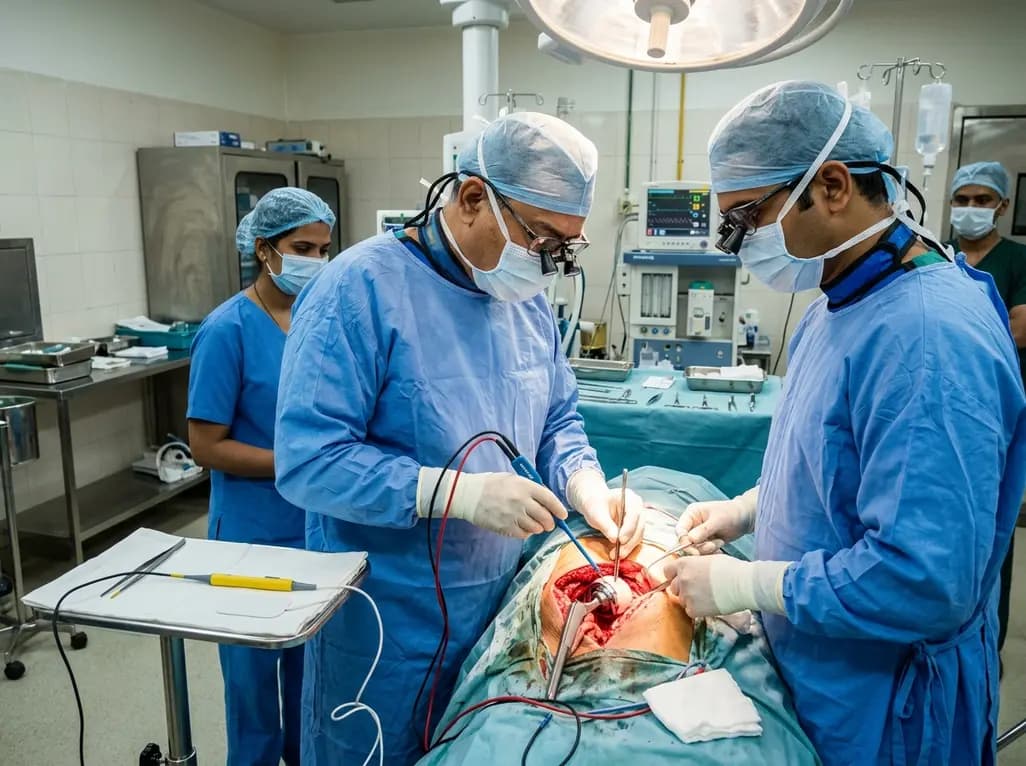
A systematic review and meta-analysis of 8 RCTs evaluated bipolar versus monopolar electrocautery in primary total hip and knee arthroplasty. Pooled data revealed no significant differences in haemoglobin drop, blood loss, or length of stay, concluding that routine bipolar electrocautery is not supported.
Yesterday

A critical review evaluates whether starch-polyphenol complexes meet structural and metabolic criteria to qualify as type 5 resistant starch. The proposed tiered evidence framework distinguishes true inclusion complexes from simple mixtures, clarifying mechanisms for glycemic modulation and gut fermentation.
Today
