.webp&w=384&q=75)

Loading, please wait...
Loading, please wait...

Spontaneous intracranial hypotension typically presents with orthostatic headaches, nausea, neck stiffness, and vestibular complaints. However, persistent extradural fluid leakages can occasionally lead to devastating motor complications. Among the most debilitating sequelae is bibrachial amyotrophy, a clinical syndrome characterized by bilateral upper extremity motor weakness and wasting with relative preservation of lower extremity and bulbar musculature. Because its clinical presentation mimics primary motor neuron diseases, clinicians often fail to identify the underlying cerebrospinal fluid leak promptly.
Historically, researchers considered upper limb wasting in cerebrospinal fluid hypovolemia an exceptionally rare and poorly documented phenomenon. Consequently, physicians frequently misdiagnose affected patients with incurable disorders such as amyotrophic lateral sclerosis, cervical spondylotic myelopathy, or Hirayama disease. Nevertheless, mounting evidence shows that recognizing this distinct manifestation early is essential. Unlike degenerative neuromuscular illnesses, weakness arising from dural tears can often be arrested or improved with targeted spine interventions.
To clarify the distinct clinical, demographic, and neuroradiological hallmarks of this secondary neuromuscular deficit, investigator Wouter I. Schievink and colleagues conducted an extensive case-control investigation. Their findings provide critical diagnostic clarity for neurologists, radiologists, and spine specialists evaluating unexplained progressive upper limb atrophy.
The investigators examined a cohort of 25 consecutive patients presenting with spontaneous intracranial hypotension complicated by bibrachial amyotrophy at a high-volume quaternary referral center between 2001 and 2024. For rigorous comparison, the team matched each patient with two control individuals experiencing spontaneous spinal cerebrospinal fluid leaks without arm amyotrophy. The controls were closely matched according to the specific anatomical site and nature of the underlying dural leak, separating ventral dural tears from lateral meningeal diverticula, as well as the calendar year of evaluation.
Before receiving a definitive diagnosis, patients in the amyotrophy cohort experienced neurological deficits for an average duration of 51 months. This prolonged latency period highlights the profound diagnostic challenges associated with non-headache presentations of intracranial hypotension. Moreover, demographic evaluation revealed striking differences between cohorts. Although spontaneous intracranial hypotension generally shows a noticeable female predilection in the wider population, men were vastly overrepresented among individuals developing upper limb amyotrophy, accounting for 80 percent of cases compared with merely 18 percent in the matched control group.
This remarkable male preponderance suggests that mechanical factors, occupational physical strain, differences in spinal column geometry, or dural elasticity may alter susceptibility to anterior spinal cord distortion in men. Therefore, clinicians must maintain high suspicion when adult male patients present with painless bilateral arm weakness alongside atypical spinal fluid collections.
The pathophysiological mechanism causing selective anterior horn motor damage in spontaneous spinal leaks is complex yet identifiable on neuroimaging. Large, chronic extradural cerebrospinal fluid collections—particularly those situated longitudinally along the ventral aspect of the cervical and upper thoracic canal—generate sustained mass effect and mechanical traction. In addition, persistent low intraspinal pressure alters local venous hemodynamics and impairs microvascular perfusion to the anterior spinal artery territory.
This persistent hypoperfusion and physical compression preferentially injure the vulnerable anterior horn cells situated within the cervical enlargement (C5 through T1 levels). Consequently, magnetic resonance imaging frequently demonstrates symmetric, bilateral T2-hyperintense intramedullary lesions within the anterior horns, often termed the classic owl-eyes sign. Furthermore, prominent ventral longitudinal epidural fluid collections and dorsal spinal cord displacement are hallmark radiographic markers.
Importantly, conventional magnetic resonance imaging of the brain in these patients may display subtle or even absent classic intracranial features of intracranial hypotension, such as pachymeningeal enhancement or tonsillar herniation. The acephalgic nature of some chronic leaks frequently obscures the clinical picture. Hence, dynamic computed tomography myelography, digital subtraction myelography, or specialized spine magnetic resonance hydrography are crucial to localize the causative dural breach.
Distinguishing bibrachial amyotrophy secondary to cerebrospinal fluid leakage from primary amyotrophic lateral sclerosis represents a high-stakes clinical challenge. Both conditions can present with insidiously progressive upper extremity paresis, pronounced muscle wasting of the deltoids, biceps, or intrinsic hand musculature, and active fasciculations. Furthermore, electromyography in both entities shows chronic neurogenic changes and active denervation potentials in multiple cervical myotomes.
However, key features help clinicians separate this mechanical myelopathy from rapidly fatal neurodegeneration. Patients with dural tears often lack lower extremity upper motor neuron signs, true bulbar involvement, and rapid respiratory decompensation during the early years of disease. Additionally, a detailed medical history may uncover a prior orthostatic headache that gradually transformed into a dull, non-positional ache or completely resolved as epidural collections expanded.
Whenever an imaging study reveals bilateral anterior horn signal abnormality accompanied by ventral extradural fluid, physicians must never assign a diagnosis of motor neuron disease without systematically excluding an occult dural rupture. Overlooking this distinction deprives patients of potentially curative or stabilizing intervention.
Management of this condition focuses on closing the underlying spinal defect to halt extradural fluid accumulation and restore physiologic cerebrospinal fluid dynamics. Depending on the precise anatomic configuration and leak rate, interventions range from targeted epidural blood patching with autologous blood and fibrin glue to microsurgical primary repair or clip ligation of dural tears.
The study demonstrated that definitive closure of the spinal breach prevents progressive neurological deterioration. In many patients, repairing the dural defect stabilizes motor deficits, and several individuals achieve meaningful functional recovery of upper extremity power, even after years of active symptoms. This recovery contrasts sharply with the inevitable, rapid downhill course observed in degenerative motor neuron disorders.
Nevertheless, long-standing anterior horn cell ischemia can leave permanent residual weakness or localized atrophy if surgical correction occurs too late. Consequently, prompt diagnostic referral, precise myelographic localization, and definitive repair by experienced multidisciplinary teams are paramount to maximizing clinical recovery and mitigating lifelong disability.
Disclaimer: This content is for informational and educational purposes only. It is not intended to be a substitute for professional medical advice, diagnosis, or treatment. Always seek the advice of your physician or other qualified health provider with any questions you may have regarding a medical condition or treatment. Never disregard professional medical advice or delay in seeking it because of something you have read here. Refer to the latest local and national guidelines for clinical practice.
References
It is a rare motor complication of chronic spontaneous intracranial hypotension where persistent ventral extradural cerebrospinal fluid collections cause mechanical compression and ischemia of cervical anterior horn cells. This process leads to progressive, symmetric muscle weakness and prominent wasting confined primarily to both upper limbs.
The condition closely mimics amyotrophic lateral sclerosis because both entities cause painless, progressive upper limb weakness, muscular atrophy, and fasciculations with chronic denervation on electromyography. Without careful spine imaging looking for ventral fluid collections, clinicians may fail to recognize the treatable mechanical leak.
Yes. Targeted epidural patching or surgical closure of the dural tear reliably halts disease progression in most patients. Furthermore, many individuals experience substantial motor recovery and functional improvement, especially when intervention occurs before irreversible cervical anterior horn cell necrosis has developed.

Read summarized clinical updates, watch expert medical content, and earn CME certifications right from your smartphone.
Open your smartphone camera to scan and install.





A landmark case-control study reveals key characteristics of bibrachial amyotrophy in patients with spontaneous intracranial hypotension, emphasizing male predominance, extensive ventral spinal CSF collections, and the diagnostic necessity of distinguishing this reversible entity from amyotrophic lateral sclerosis.
Today
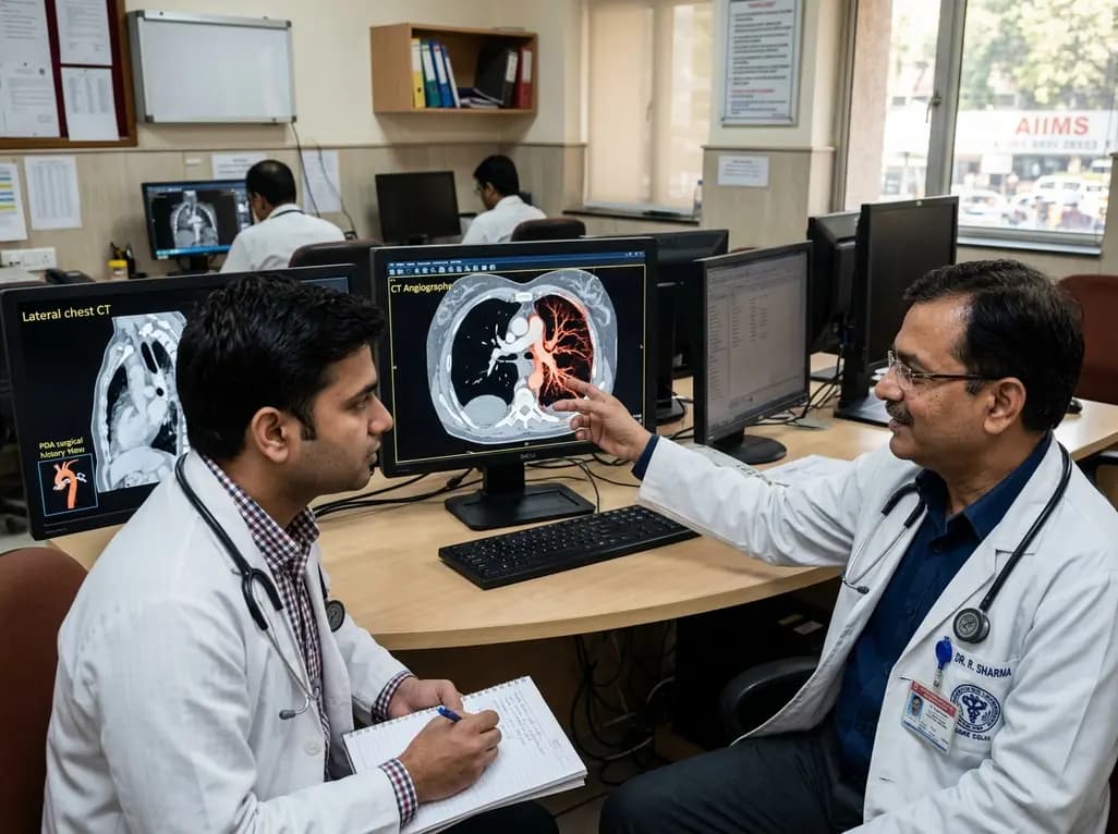
Congenital unilateral absence of the pulmonary artery can remain undetected until adulthood. This case report examines a young male presenting with hemoptysis after prior PDA surgery, highlighting the critical role of CT angiography in mapping systemic collaterals and guiding appropriate clinical intervention.
Today

Perimenopausal obstructive sleep apnea often goes undetected due to atypical polysomnographic presentations and symptom overlap with menopause. This review details its neurocognitive mechanisms, polysomnography markers, and precision management strategies.
Today

Mitochondrial deubiquitinases regulate organelle turnover by editing ubiquitin tags on damaged mitochondria. Discover how targeting these enzymes, notably USP30, offers therapeutic strategies for neurodegenerative, cardiovascular, and metabolic disorders.
Today

A rare case of a pediatric pathological femoral fracture caused by occult telangiectatic osteosarcoma demonstrates the risks of misdiagnosis after trauma, where intramedullary nailing prompted iatrogenic tumor dissemination and required modular total endoprosthetic reconstruction.
Today

A comprehensive analysis of DRCRnet studies reveals that 15.1% of participants discontinue diabetic retinopathy clinical trials. Identifying baseline demographic and clinical predictors helps ophthalmologists and endocrinologists mitigate real-world treatment dropouts and protect long-term visual outcomes.
Today
