.webp&w=384&q=75)

Loading, please wait...
Loading, please wait...

Spatial neglect represents one of the most debilitating neurocognitive complications following a cerebrovascular accident, particularly after right-hemispheric stroke. While clinicians historically characterize neglect as an isolated visual impairment, emerging evidence demonstrates that multimodal deficits frequently co-occur. Consequently, neurorehabilitation researchers have increasingly explored cross-modal sensory interventions to enhance patient recovery. In particular, the application of auditory cueing in spatial neglect has garnered notable interest as a non-invasive therapeutic approach. However, recent clinical investigations reveal that the therapeutic success of acoustic stimuli varies widely across patient populations. Clinicians must understand how underlying auditory spatial processing directly dictates the efficacy of these cross-modal interventions.
Hemispatial neglect impairs a patient's ability to orient attention, detect stimuli, and respond to environmental events occurring within the contralesional space. Although bedside assessments predominantly evaluate visual exploration through cancellation tests and line bisection, neglect reflects a broader supramodal network breakdown. Patients often demonstrate concurrent tactile and acoustic deficits that compromise functional recovery. When auditory orienting deteriorates alongside vision, patients cannot localize sound sources accurately within their neglected hemifield. Furthermore, this impairment undermines basic daily activities, such as navigating busy urban environments or engaging in group conversations. In India, where post-stroke rehabilitation services often face heavy patient loads, identifying specific sensory profiles becomes essential for optimizing recovery. Many neurorehabilitation protocols assume that preserved auditory channels can automatically compensate for defective visual attention. Nevertheless, clinicians frequently overlook subclinical auditory deficits during standard evaluations. When brain damage disrupts multimodal integration pathways, presenting auditory signals may not trigger the intended visual reorientation. Consequently, treating neglect as a purely visual disorder prevents therapists from delivering personalized and efficacious rehabilitation. Recognizing neglect as a multidimensional condition enables clinicians to stratify stroke survivors far more accurately.
Cross-modal sensory cueing operates on the principle that salient inputs in one sensory modality can exogenously capture attention and direct gaze toward neglected sectors. Therefore, clinicians frequently introduce acoustic signals to guide spatial realignment toward the left hemifield. A recent landmark study investigated this paradigm by evaluating sixty participants, including healthy controls and right-hemisphere stroke survivors with or without visual neglect. The researchers rigorously stratified visual neglect patients based on their auditory spatial localisation performance. Participants completed computer-based visual search tasks across four experimental sound conditions: congruent spatial cues, incongruent spatial cues, non-spatial monophonic sounds, and silent baseline trials. Strikingly, the therapeutic benefit of congruent sound cues emerged exclusively in patients with preserved auditory localisation capacity. Individuals presenting with combined visual and auditory neglect failed to achieve meaningful visual search acceleration. Moreover, overall sound localisation accuracy showed a significant positive correlation with visual search speed during congruent presentations. Thus, auditory cueing in spatial neglect does not provide an indiscriminate, universal benefit across all stroke survivors. Instead, the intervention requires functional auditory spatial processing networks to effectively cross-cue the visual system.
To elucidate the structural substrates underlying these clinical differences, investigators conducted voxel-based lesion-symptom mapping among the stroke cohort. This advanced neuroimaging analysis demonstrated that impaired auditory spatial localisation correlates strongly with focal disruptions in the right superior longitudinal fasciculus. Specifically, damage centered on the first and second branches of this major white matter tract. The superior longitudinal fasciculus constitutes a critical frontoparietal pathway that coordinates attentional control, sensorimotor integration, and spatial coordinate mapping across the cerebral hemispheres. Consequently, when an ischemic or hemorrhagic lesion disconnects these subcomponents, the brain loses the structural scaffolding necessary for sound-guided spatial attention. Stroke patients with intact tracts preserve their capacity to compute auditory spatial coordinates, allowing sound to prime visual saccades toward neglected sectors. Conversely, extensive structural disruption across these white matter bundles abolishes cross-modal transfer. Neurologists should therefore scrutinize structural MRI scans for superior longitudinal fasciculus involvement when planning rehabilitation trajectories. Recognizing tract integrity provides valuable prognostic insight into whether a patient can benefit from acoustic guidance strategies.
These neuroscientific observations carry profound implications for routine stroke assessment and rehabilitation planning. Standard clinical batteries, such as the Behavioral Inattention Test, concentrate almost exclusively on visuospatial pen-and-paper tasks. Consequently, healthcare providers frequently miss co-occurring auditory spatial deficits in acute and subacute stroke survivors. Clinicians must integrate systematic auditory localisation tests into their routine neurofunctional examinations. Simple dichotic listening paradigms, spatialised sound presentations, and bedside auditory localisation arrays can rapidly uncover hidden deficits. Furthermore, identifying patients with isolated visual neglect allows therapists to deploy auditory cueing protocols with high confidence. Conversely, identifying combined visual-auditory neglect prevents clinicians from wasting precious rehabilitation hours on ineffective acoustic strategies. In resource-conscious clinical environments, such targeted screening streamlines therapy allocation and improves functional outcomes. Additionally, speech therapists and occupational therapists can align their interventions once they understand the exact sensory phenotype. Moving beyond single-modality assessments guarantees that neurorehabilitation strategies match the biological reality of individual patient lesions.
Modern neurorehabilitation is steadily advancing away from generic, one-size-fits-all therapy toward precision medicine paradigms. For patients with preserved auditory processing, digital health developers can design targeted wearable devices that deliver congruent spatial acoustic alerts during everyday navigation. Similarly, immersive virtual reality platforms can combine spatialised audio with dynamic visual exploration tasks to accelerate recovery. Meanwhile, patients suffering from multimodal neglect require alternative sensory compensatory pathways. Clinicians can explore galvanic vestibular stimulation, neck muscle vibration, or limb activation training to bypass damaged frontoparietal auditory networks. Furthermore, non-invasive neuromodulation techniques, such as repetitive transcranial magnetic stimulation or transcranial direct current stimulation, may help restore balanced interhemispheric attention. Integrating these technologies into standard hospital protocols will substantially elevate the standard of care for stroke survivors. Ultimately, rigorous multimodal characterization enables multidisciplinary care teams to deliver customized therapies that significantly reduce long-term disability and foster independence.
Auditory cueing in spatial neglect uses spatialised acoustic stimuli to attract attention toward the neglected contralesional side. By delivering sounds from specific locations, therapists trigger exogenous attentional orienting. This sensory cue primes the visual system, helping stroke survivors initiate saccades and locate objects faster in their impaired visual field.
Patients fail to benefit from auditory cueing when strokes damage shared frontoparietal spatial networks, causing concurrent auditory neglect. If individuals cannot accurately localize sound sources, cross-modal priming mechanisms break down completely. Consequently, auditory cues cannot guide visual attention unless white matter tracts supporting sound localisation remain structurally functional.
Auditory spatial performance depends heavily on the right superior longitudinal fasciculus, particularly its first and second branches. Lesions affecting these white matter tracts disrupt vital communication between frontal and parietal attention regions. This anatomical disconnection prevents accurate contralesional sound localization, precluding cross-modal sensory recovery in hemispatial neglect.
Disclaimer: This content is for informational and educational purposes only... Refer to the latest local and national guidelines for clinical practice.
References

Read summarized clinical updates, watch expert medical content, and earn CME certifications right from your smartphone.
Open your smartphone camera to scan and install.





A novel study highlights that cross-modal auditory cueing in spatial neglect benefits stroke patients only when auditory spatial localisation remains intact. Lesions in the right superior longitudinal fasciculus explain auditory deficits, emphasizing multimodal testing for tailored neurorehabilitation.
Today
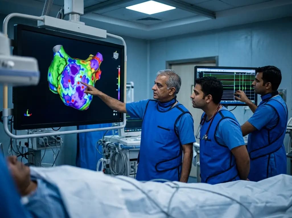
A study of 1,428 AF ablation patients reveals that over 60% of left atrial non-PV triggers arise within low-voltage areas, with anterior and roof triggers colocalizing entirely within fibrotic substrate. Identifying and ablating these sites is critical for long-term arrhythmia freedom.
Today

A comprehensive meta-analysis of over 278,000 women demonstrates that maternal atopic dermatitis significantly increases risks of premature rupture of membranes and low birth weight. Discover essential clinical insights on disease mechanisms, serial antenatal growth monitoring, and safe therapeutic interventions.
Today

A pioneering study highlights the first United States experience using the FARAPOINT focal pulsed-field ablation catheter for refractory ventricular tachycardia. The catheter achieved complete acute non-inducibility across tested patients without procedural complications, marking a milestone in ventricular ablation.
Today

A biomechanical study shows that augmenting thin fascia lata grafts with a reinforced polyester patch doubles ultimate tensile strength and stiffness, matching the performance of thick grafts for superior capsule reconstruction while reducing donor-site morbidity.
Today

A cross-sectional study reveals that elevated METS-IR is independently associated with higher myopia odds in adolescents, showing biological synergy with obesity and highlighting the need for metabolic screening in pediatric eye care.
Today
