.webp&w=384&q=75)

Loading, please wait...
Loading, please wait...
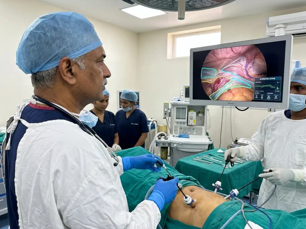
Surgical robotics and computer vision continue to transform minimally invasive surgery, providing clinicians with unprecedented intraoperative guidance. In particular, laparoscopic inguinal hernia repair via the transabdominal preperitoneal (TAPP) approach demands meticulous visual identification of complex anatomical planes. Surgeons must reliably isolate loose connective tissue while preserving crucial structures such as the vas deferens, testicular vessels, and regional neural pathways. Consequently, any cognitive ambiguity during tissue dissection can lengthen operative duration and elevate the risk of iatrogenic injury. Recent advances in deep learning offer promising solutions by processing high-definition endoscopic video streams in real time. By segmenting anatomical boundaries and highlighting dissection planes, computer vision systems act as an intelligent co-pilot in the operating theater. A groundbreaking comparative study evaluated the clinical integration of this technology during laparoscopic groin hernia repairs. The findings highlight how real-time artificial intelligence navigation (RAIN) enhances procedural efficiency without compromising patient safety, establishing a compelling case for smart surgical navigation.
The success of preperitoneal groin hernia repair depends on accurate plane identification across the myopectoral orifice. During standard TAPP procedures, surgeons navigate delicate regions including the triangle of doom and the triangle of pain. Inadvertent injury to external iliac vessels or femoral nerve branches can produce devastating patient outcomes. Furthermore, extensive scarring from chronic inflammation or previous interventions frequently obscures tissue boundaries. Consequently, identifying the exact loose connective tissue plane, often termed the dissectible layer, demands significant technical expertise. Experienced surgeons rely on subtle visual cues, such as light reflection, tissue compliance, and microvascular architecture, to guide their dissection. However, less experienced trainees often struggle with visual ambiguity, leading to hesitation and prolonged operative duration. Variations in patient anatomy, adipose tissue distribution, and prior adhesions further complicate visualization. Therefore, augmenting human visual perception with objective, algorithmic tissue mapping represents a critical development in modern surgical practice, establishing standard safety margins across diverse surgical skill levels.
To overcome anatomical visual barriers, researchers developed EUREKAα, an artificial intelligence system approved as a medical device in Japan in April 2024. The platform employs deep neural networks trained on thousands of annotated endoscopic surgical video frames from multiple academic centers. As the laparoscope captures the abdominal cavity, the software processes the incoming video feed frame-by-frame with minimal visual latency. Subsequently, the system renders a dynamic, color-coded visual overlay on a dedicated intraoperative monitor, highlighting loose connective tissue in real time. This automated layer recognition guides the surgical team directly along optimal embryonic planes. Because loose connective tissue serves as the natural boundary for dissection, visual prompts enable surgeons to progress smoothly without entering vascular or neural structures. Moreover, the secondary display provides an objective visual reference without cluttering the primary endoscopic view. Consequently, the surgical team maintains total situational awareness while referencing algorithmic guidance during critical dissection maneuvers.
To evaluate the real-world utility of artificial intelligence guidance, investigators conducted an extensive clinical study encompassing 508 consecutive TAPP procedures performed between February 2020 and December 2025. Among these cases, 54 procedures incorporated real-time AI navigation, whereas 454 cases utilized conventional laparoscopic techniques without algorithmic assistance. To mitigate treatment selection bias and balance baseline confounding variables, the authors performed rigorous propensity score matching (PSM). The matching algorithm successfully created 53 well-balanced pairs of patients, adjusting for relevant clinical parameters such as age, body mass index, hernia type, defect size, and surgeon experience level. Primary endpoints focused on total operative duration, intraoperative blood loss, and inadvertent structural injuries. Secondary endpoints analyzed postoperative recovery metrics, including wound complications, seroma formation, early recurrence, and hospital length of stay. This robust comparative design ensured a fair evaluation of AI-guided surgery against established standards.
The statistical analysis revealed notable differences in surgical efficiency between the two cohorts. In the unmatched analysis, the real-time AI navigation group demonstrated a significantly shorter mean operative time of 38.6 minutes, compared to 48.6 minutes in the conventional group. Following propensity score matching, this significant advantage persisted robustly across the cohorts. Specifically, the matched RAIN group achieved a mean operative duration of 38.5 minutes, whereas the matched conventional group required 49.2 minutes. This substantial reduction of more than ten minutes represents a meaningful improvement in operating room utilization. Importantly, this acceleration did not come at the expense of patient safety. The incidence of postoperative complications, including hematoma, urinary retention, and surgical site infections, showed no significant differences between cohorts. Thus, real-time visual assistance streamlined technical execution, eliminated unnecessary procedural hesitation, and maintained exemplary surgical safety standards throughout the study period.
The successful integration of real-time AI navigation into hernia repair signals a broader shift toward intelligent intraoperative assistance. Although EUREKAα initially targeted loose connective tissue segmentation, expanding its algorithmic capabilities to identify nerves, microvessels, and visceral boundaries will unlock greater clinical utility. Furthermore, integrating computer vision algorithms into robotic surgery consoles could allow active haptic feedback, preventing instruments from straying beyond safe anatomical boundaries. In addition to technical execution, this technology provides substantial educational value for surgical residency programs. Trainees can develop sharper anatomical intuition by comparing intraoperative views with real-time AI tissue predictions. As multicenter prospective trials validate these preliminary findings across diverse hospital environments, algorithmic surgical guidance is poised to become a routine standard. Ultimately, artificial intelligence transforms laparoscopic surgery from subjective visual interpretation into an objective, data-driven discipline that consistently elevates patient outcomes.
The artificial intelligence system analyzes intraoperative video feeds and highlights loose connective tissue planes on a dedicated monitor. Consequently, surgeons can quickly identify optimal dissection boundaries while avoiding critical nerves and blood vessels. This algorithmic guidance minimizes visual hesitation, streamlines operative workflow, and provides objective spatial orientation during complex minimally invasive dissections.
No, the clinical study demonstrated that real-time AI navigation shortened operative time without increasing postoperative complications. Propensity score-matched analyses confirmed equivalent rates of seroma, hematoma, urinary retention, and infection between AI-assisted and conventional cohorts. Therefore, algorithmic guidance improves operative efficiency while maintaining rigorous clinical safety standards.
Current commercial platforms specialize in identifying loose connective tissue layers, but subsequent software updates are actively expanding these capabilities. Developers are training models to recognize autonomic nerve trunks, the vas deferens, and microvascular structures. Future iterations will likely offer multi-structure segmentation, further enhancing surgical precision and preventing inadvertent tissue trauma.
Disclaimer: This content is for informational and educational purposes only. It is not intended to be a substitute for professional medical advice, diagnosis, or treatment. Always seek the advice of your physician or another qualified healthcare provider with any questions you may have regarding a medical condition or treatment. Refer to the latest local and national guidelines for clinical practice.
References
Mita K et al. Intraoperative real-time recognition of dissectible layers by artificial intelligence is useful in laparoscopic inguinal hernia repair. Surg Endosc. 2026 Aug 20. doi: 10.1007/s00464-026-13241-2. PMID: 42622652.
Shinohara H, et al. Real-time artificial intelligence visual support in endoscopic surgery: initial clinical evaluation of Eureka α. Surg Today. 2024;54(11):1201-1208.
Hashimoto DA, et al. Artificial intelligence in surgery: promises and perils. Ann Surg. 2018;268(1):70-76.

Read summarized clinical updates, watch expert medical content, and earn CME certifications right from your smartphone.
Open your smartphone camera to scan and install.





A landmark clinical study demonstrates that real-time AI recognition of dissectible tissue layers significantly reduces operative time without increasing complications in laparoscopic transabdominal preperitoneal (TAPP) inguinal hernia repair, marking a major milestone for surgical computer vision.
last month

A nationwide English cohort study shows that interstitial lung diseases are no longer rare, with incidence jumping 30% and 200,000 active cases. This analysis highlights clinical implications for diagnosis, multi-disciplinary management, and global health priorities across respiratory and internal medicine.
Today

A nationwide South Korean study shows underreporting of smoking among women dropped from 57.4% to 36.4% between 2008 and 2021. However, over one-third of female smokers still conceal their habit, highlighting the critical role of cotinine verification in clinical risk assessment.
Today

PepsiCo and Monster Beverage have challenged the FSSAI ban on the energy drink label in Indian courts. While companies highlight supply disruptions, regulatory actions spotlight severe cardiovascular and metabolic risks associated with excessive caffeine, sugar, and taurine consumption, especially in adolescents.
Today

A new study evaluates the rheological stability of decellularized peripheral nerve matrix hydrogels under varied storage conditions. Findings show -80°C storage and freeze-drying preserve gelation kinetics and mechanical stiffness, offering key insights for shipping and translational clinical applications.
Today

A Bayesian multilevel meta-analysis reveals that aerobic training combined with moderate carbohydrate restriction modestly lowers HbA1c in type 2 diabetes. However, sparse data and very low certainty leave incremental benefits over exercise or diet alone unproven, highlighting the need for individualized care.
Yesterday
