

Loading, please wait...
Loading, please wait...

Modern cardiovascular medicine increasingly relies on innovative AI-based CT-FFR technology to evaluate stable chest pain and detect myocardial ischemia non-invasively. Coronary computed tomography angiography provides exceptional anatomical visualization of luminal narrowings. However, anatomical assessment alone frequently fails to determine whether an intermediate stenosis produces hemodynamic ischemia. Consequently, clinicians often perform invasive coronary angiography to measure fractional flow reserve directly with pressure wires. This invasive pathway introduces procedure-related risks, elevates healthcare expenditures, and often reveals non-obstructive disease.
To resolve this diagnostic limitation, researchers developed novel deep learning algorithms. Specifically, these computational platforms rapidly calculate lesion-specific physiology directly from standard tomographic datasets. The algorithms extract vascular geometry and simulate fluid mechanics within seconds without requiring off-site supercomputing facilities. Furthermore, point-of-care solutions like CorEx enable on-site functional evaluations directly within the diagnostic radiology suite. Therefore, clinicians can rapidly differentiate hemodynamically significant lesions from functionally benign narrowings during initial outpatient triage. This breakthrough substantially enhances diagnostic confidence while streamlining downstream catheterization referrals.
Recent clinical investigations evaluated how patient selection and baseline disease prevalence influence algorithmic accuracy. In the prospective PACIFIC-1 study cohort, researchers analyzed two hundred and eight patients across six hundred and twelve coronary vessels. Notably, all participants underwent comprehensive invasive pressure wire interrogations regardless of their initial computed tomography findings. In this unselected cohort, the AI platform demonstrated an overall per-vessel diagnostic accuracy of seventy-seven percent.
Meanwhile, standard anatomical stenosis thresholds of fifty and seventy percent achieved seventy-six and seventy-four percent accuracy, respectively. Statistical comparisons confirmed comparable diagnostic utility between the functional software and anatomical grading in this protocol-driven cohort. In contrast, investigators observed markedly different diagnostic patterns when examining a consecutive real-world clinical cohort. This real-world cohort comprised two hundred and two patients presenting with severe anatomic stenosis exceeding seventy percent.
Under these high-risk conditions, the deep learning model achieved a per-vessel accuracy of sixty-three percent. Furthermore, the model significantly outperformed conventional anatomical stenosis thresholds of fifty and seventy percent. Thus, algorithmic functional assessment provides superior discriminatory power in heavily diseased patient populations presenting for routine invasive coronary evaluation.
Discrepancies between anatomical narrowing and ischemic physiology represent a persistent clinical dilemma in interventional cardiology. Visual assessment on tomographic scans often overestimates the hemodynamic impact of intermediate and calcified lesions. For instance, dense coronary calcification produces substantial blooming artifacts that obscure the true vascular lumen. Consequently, anatomical interpretation alone precipitates numerous diagnostic angiograms that ultimately uncover non-ischemic physiology. The deep learning software overcomes these visual hurdles by applying complex non-linear hemodynamic predictions.
Moreover, the neural algorithm analyzes lumen tapering, lesion length, and subtended myocardial mass simultaneously. In the real-world registry, anatomical thresholds of fifty and seventy percent yielded diagnostic accuracies of only fifty-two and fifty-seven percent. The artificial intelligence model surpassed both anatomic parameters by delivering a sixty-three percent accuracy rate. Therefore, integrating functional simulation prevents premature catheterization referrals in patients with anatomically severe yet hemodynamically non-limiting disease.
In addition, this physiological precision preserves catheterization laboratory capacity for patients who genuinely require coronary revascularization. Clinicians thereby optimize invasive procedural yields while protecting lower-risk individuals from unnecessary arterial instrumentation.
Observational cohorts in cardiovascular medicine inherently reflect referral bias because symptomatic patients undergo prioritized advanced diagnostics. When cardiologists suspect critical disease, they preferentially refer patients with severe lesions for invasive catheterization. As a result, clinical registries naturally accumulate higher proportions of complex multivessel disease and calcified plaques. This verification bias alters pre-test probability and directly depresses overall diagnostic specificity.
Nevertheless, the artificial intelligence model maintained consistent performance despite these demanding clinical conditions. The software processed coronary geometries autonomously without human editing or off-site core laboratory delays. Furthermore, this point-of-care capability enables emergency departments and outpatient clinics to formulate rapid therapeutic strategies. Traditional computational fluid dynamics often require hours of processing time on remote server clusters.
In comparison, neural networks deliver functional assessments in less than one minute. Consequently, clinicians can integrate ischemic modeling directly into daily diagnostic workflows without postponing patient care. This operational efficiency represents a transformative technological advancement for high-throughput cardiovascular imaging centers worldwide.
Coronary artery disease exhibits a premature, aggressive, and highly diffuse clinical phenotype across Indian populations. Indian patients frequently present with extensive multivessel involvement, elevated calcification burdens, and smaller reference vessel diameters. In addition, high out-of-pocket healthcare expenditures impose substantial economic stress on families across the country. Unnecessary diagnostic catheterizations consume scarce resources and expose patients to potential vascular access complications. Therefore, implementing non-invasive functional imaging offers immense clinical and economic advantages in Indian healthcare settings.
By deploying on-site algorithmic platforms, tertiary and tier-two cardiac centers can accurately triage symptomatic individuals. Notably, functional triage ensures that only patients with verified ischemic physiology proceed to the catheterization laboratory. Meanwhile, physicians can safely direct patients with non-ischemic intermediate stenoses toward aggressive medical therapy and intensive lifestyle modification. This targeted allocation minimizes healthcare expenditures while maximizing clinical outcomes for millions of vulnerable cardiac patients.
Furthermore, widespread adoption of automated functional modeling will democratize advanced cardiovascular diagnostics beyond metropolitan centers. Indian healthcare institutions must therefore establish standardized acquisition protocols to maximize tomographic image quality and diagnostic reliability.
Standard coronary computed tomography angiography provides detailed anatomical visualization of plaque and luminal narrowing. However, it cannot reliably identify whether a moderate stenosis impedes myocardial blood flow. In contrast, AI-based CT-FFR applies deep learning algorithms to predict coronary hemodynamics directly from tomographic datasets. Consequently, it effectively identifies lesion-specific ischemia without invasive pressure wire instrumentation. This functional evaluation significantly reduces unnecessary diagnostic catheterizations while guiding appropriate revascularization decisions.
Baseline disease prevalence profoundly influences the clinical utility of machine learning hemodynamic software. In low-to-intermediate prevalence cohorts, high negative predictive value enables confident exclusion of obstructive ischemia. Conversely, in real-world cohorts with high disease prevalence and severe anatomic stenosis, visual grading frequently overestimates hemodynamic significance. Under these demanding conditions, artificial intelligence maintains superior diagnostic accuracy over anatomical thresholds alone. Therefore, algorithmic physiological assessment helps clinicians avoid inappropriate interventions in heavily diseased populations.
Clinicians can safely defer invasive catheterization in stable patients when functional assessment reveals non-ischemic values exceeding 0.80. Substantial clinical evidence confirms that deferring intervention for non-ischemic lesions carries very low adverse cardiac event rates. Furthermore, deferred patients achieve excellent outcomes under guideline-directed medical therapy. However, physicians must evaluate image quality, symptom severity, and anatomical high-risk plaque characteristics carefully. When high image quality and negative functional values align, deferring invasive procedures represents a safe, guideline-supported clinical approach.
Disclaimer: This content is for informational and educational purposes only... Refer to the latest local and national guidelines for clinical practice.
References
Coerkamp CF et al. Performance of artificial intelligence-based coronary CT angiography-derived fractional flow reserve across cohorts with different prevalence of obstructive coronary artery disease. J Cardiovasc Comput Tomogr. 2026 Oct 19. doi: undefined. PMID: 42897082.
Peters B, Paul JF, Symons R, Franssen WMA, Nchimi A, Ghekiere O. Invasive fractional-flow-reserve prediction by coronary CT angiography using artificial intelligence vs. computational fluid dynamics software in intermediate-grade stenosis. Int J Cardiovasc Imaging. 2024;40(9):1875-1880.
Kloth C, Brendel JM, Kübler J, et al. CT-FFR: How a new technology could transform cardiovascular diagnostic imaging. Rofo. 2025;197(5):482-491.

Read summarized clinical updates, watch expert medical content, and earn CME certifications right from your smartphone.
Open your smartphone camera to scan and install.





A multicenter evaluation reveals that AI-based CT-FFR outperforms anatomical stenosis grading in high-prevalence cohorts, offering rapid non-invasive ischemia detection to guide revascularization and reduce unnecessary invasive coronary angiography procedures.
Today

The Food Safety and Standards Authority of India has issued an urgent advisory warning food business operators against flouting mandatory labelling rules and cautionary warnings on botanical extracts. Learn how these regulatory actions impact clinical safety, patient discussions, and supplement surveillance in India.
Today
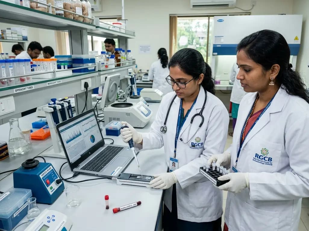
Biotechnology researchers in India are developing an automated liquid biopsy platform to detect cervical malignancy from blood samples. Supported by the MAHA MedTech Mission, this initiative applies nanopore sequencing and piRNA biomarker detection to eliminate invasive speculum exams and expand rural screening.
Today
A systematic review and meta-analysis of 8 RCTs evaluated bipolar versus monopolar electrocautery in primary total hip and knee arthroplasty. Pooled data revealed no significant differences in haemoglobin drop, blood loss, or length of stay, concluding that routine bipolar electrocautery is not supported.
Today
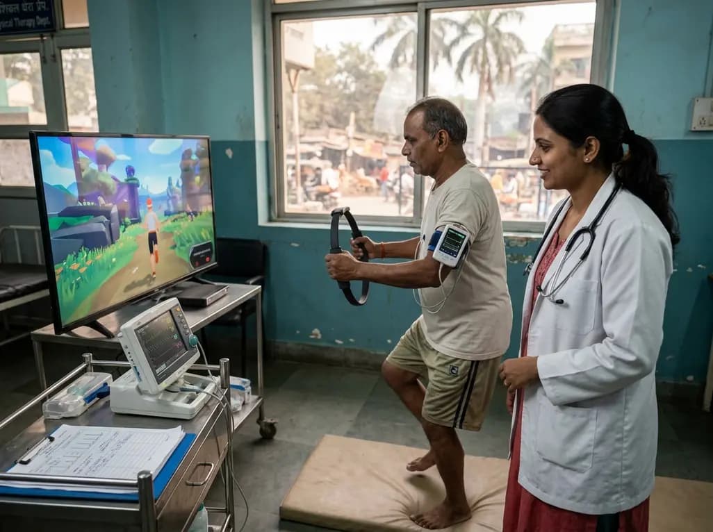
A clinical study evaluated Ring Fit Adventure in adults with chronic conditions, revealing that exergaming achieves moderate-to-vigorous exercise intensity while maintaining hemodynamic stability, high enjoyment, and zero adverse events.
Today
