.webp&w=384&q=75)

Loading, please wait...
Loading, please wait...
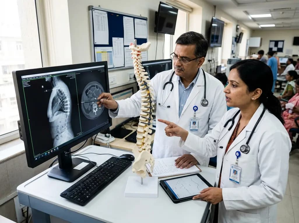
Adult spinal deformity surgery often requires extensive posterior instrumentation to restore sagittal balance and correct severe mechanical alignment deficits. However, complex reconstructive procedures carry significant risks of structural complications at adjacent anatomical levels. Among these complications, proximal junctional failure represents a major clinical challenge for modern reconstructive spine surgeons. Anterior column realignment has emerged as a powerful technique enabling major focal sagittal lordosis correction. Nevertheless, achieving substantial angular correction across anterior disc spaces can place excessive biomechanical stress on the upper instrumented vertebra. Consequently, acute proximal junctional failure can develop within six months after major spinal surgery, often requiring secondary revision procedures. Recent clinical investigation provides crucial insights into the overall incidence and specific risk factors associated with this severe postoperative outcome. Understanding these anatomical and technical predictors allows surgeons to refine surgical planning, optimize construct design, and improve long-term clinical safety for adult spinal deformity patients.
Evaluating mechanical complications after anterior column realignment requires distinguishing between structural failure mechanisms, specifically bony fractures and soft tissue disruption. Clinical research indicates that proximal junctional failure occurs in roughly thirty-one percent of patients undergoing complex anterior column realignment for severe sagittal imbalance. Furthermore, nearly thirty-nine percent of patients affected by acute proximal junctional failure ultimately require surgical revision within six months. When analyzing specific failure modes, bony structural compromises, such as vertebral body fractures or screw pullout, account for the vast majority of revision cases. Conversely, soft tissue failures involving ligamentous disruption represent a smaller proportion of reoperations. The higher frequency of bony failure highlights the profound biomechanical strain transmitted to the uppermost instrumented vertebra following aggressive anterior lordotic correction. Therefore, spine specialists must focus on bone density enhancement and biomechanical load distribution when planning extensive adult deformity corrections.
Radiographic assessment plays a central role in identifying patients at elevated risk for mechanical complications following complex spine realignment. Clinical studies highlight several key radiographic parameters that strongly correlate with the need for revision surgery. Notably, the cranial screw angle at the upper instrumented vertebra serves as an independent predictor of revision due to acute proximal junctional failure. Steep trajectory angles at construct termination create severe stress concentrations at the bone-screw interface, predisposing the upper vertebra to catastrophic fracture or fixation pullout. Additionally, preoperative thoracic kyphosis, postoperative thoracic kyphosis, and the immediate postoperative proximal junctional angle significantly influence structural stability. When surgeons achieve substantial lordotic corrections anteriorly without adequately accounting for adjacent thoracic compensation, proximal junctional stress escalates rapidly. Consequently, meticulous pre-operative planning must incorporate careful sagittal alignment targets that accommodate natural regional curvature and spinal flexibility.
To minimize revision rates associated with acute proximal junctional failure, surgical techniques must adapt to reduce stress concentrations at the construct transition zone. Implementing soft tissue preservation protocols at the upper instrumented vertebra represents an essential strategy for maintaining junctional stability. Protecting supraspinous and interspinous ligaments preserves crucial posterior tension bands that cushion mechanical forces during flexion. Additionally, adopting biomechanical strategies such as transitional instrumentation, transverse process hooks, or flexible tethering can effectively soften the abrupt stiffness gradient between fused and mobile spinal segments. Prophylactic cement augmentation at the uppermost instrumented level also offers substantial protection against acute vertebral body collapse, particularly in elderly patients with compromised bone mineral density. Surgeons must carefully balance aggressive anterior column realignment with prudent posterior stabilization strategies, utilizing intraoperative image guidance to optimize screw placement angles and minimize cortical strain.
Managing adult spinal deformity requires a comprehensive approach integrating clinical assessment, advanced imaging, and tailored surgical execution. Because acute proximal junctional failure often presents within the first few months following anterior column realignment, close clinical surveillance during the early postoperative phase is essential. Care teams must maintain high suspicion when patients report acute localized back pain or demonstrate progressive sagittal decompensation. Early radiographic evaluation allows prompt identification of structural failure, facilitating timely intervention before severe neurological deficits occur. Furthermore, optimizing preoperative bone health through targeted medical therapies, such as osteoanabolic agents, can significantly improve bone quality and construct fixation strength. Collaborative perioperative care involving endocrinologists, physical therapists, and spine specialists ensures optimal medical optimization and post-surgical rehabilitation. Ultimately, recognizing key risk factors empowers clinicians to deliver safer, individualized surgical care.
Improving long-term clinical outcomes in adult spinal deformity surgery hinges on minimizing avoidable mechanical complications that necessitate secondary revision. While anterior column realignment offers exceptional restoration of lordosis, mitigating junctional stress remains critical to overall surgical success. Future research must focus on prospective clinical trials evaluating dynamic stabilization systems, optimized screw trajectories, and patient-specific alignment formulas. Additionally, advanced artificial intelligence modeling may soon assist spine surgeons in predicting individualized proximal junctional stress distributions before entering the operating room. By combining predictive analytics with established risk parameters like cranial screw angle and regional kyphosis, surgical teams can customize surgical approaches for every unique deformity. Standardizing postoperative follow-up protocols ensures consistent monitoring of junctional angles, allowing early biomechanical intervention when minor subluxation appears.
Acute proximal junctional failure is a severe mechanical complication occurring within six months after adult spinal deformity correction. It involves structural compromise at or near the top of the surgical construct, including vertebral body fractures, implant dislocation, soft tissue disruption, or progressive angular deformity. This acute breakdown frequently causes severe pain, mechanical instability, or neurological symptoms, often requiring secondary revision surgery to restore structural integrity and relieve nerve compression.
Anterior column realignment enables significant lordotic correction across disc spaces, effectively restoring sagittal balance in severe spinal deformities. However, rapidly inducing major focal angular correction increases mechanical leverage and stress concentration at the upper instrumented vertebra. Consequently, if posterior stabilization or adjacent thoracic alignment is not perfectly matched, excessive forces can overload terminal anchors, leading to acute structural breakdown or vertebral body fracture shortly after surgery.
Key risk factors for revision surgery include a steep cranial screw angle at the upper instrumented vertebra, severe preoperative thoracic kyphosis, increased postoperative thoracic kyphosis, and an elevated immediate proximal junctional angle. Furthermore, bony structural failures, such as vertebral fractures, carry a significantly higher likelihood of requiring revision compared to soft tissue failures. Identifying these parameters allows surgeons to adjust anchor angles and employ prophylactic reinforcement techniques.
Disclaimer: This content is for informational and educational purposes only and does not constitute professional medical advice, diagnosis, or treatment. Always seek the advice of a qualified healthcare provider with any questions regarding a medical condition. Refer to the latest local and national guidelines for clinical practice.
References
Park JS et al. Incidence of and risk factors for revision surgery due to acute proximal junctional failure after anterior column realignment for adult spinal deformity. J Neurosurg Spine. 2026 Aug 07. doi: 10.3171/2026.3.SPINE25135. PMID: 42566791.

Read summarized clinical updates, watch expert medical content, and earn CME certifications right from your smartphone.
Open your smartphone camera to scan and install.





A clinical study evaluates acute proximal junctional failure and revision risk factors following anterior column realignment for adult spinal deformity. Cranial screw angle at UIV and thoracic kyphosis emerge as major independent predictors, emphasizing technical refinements to prevent revision surgery.
last month

A study verifies the quantitative risk assessment model using EC1.6 values from LLNA: BrdU-ELISA to predict skin sensitization from medical devices. Comparing FreeStyle Libre and Libre 2, researchers confirmed how reducing isobornyl acrylate below acceptable levels prevents allergic contact dermatitis.
Today

A recent longitudinal study evaluated the link between prenatal loss of control eating and cardiovascular health using Life's Essential 8 frameworks. While direct associations across pregnancy were non-significant, the high prevalence of dysregulated eating highlights critical implications for obstetric practice.
Today
Explore modern perioperative management protocols for clinical xenotransplantation. Learn how costimulation blockade, anti-inflammatory perfusion, and coagulopathy rescue overcome interspecies hurdles.
Today
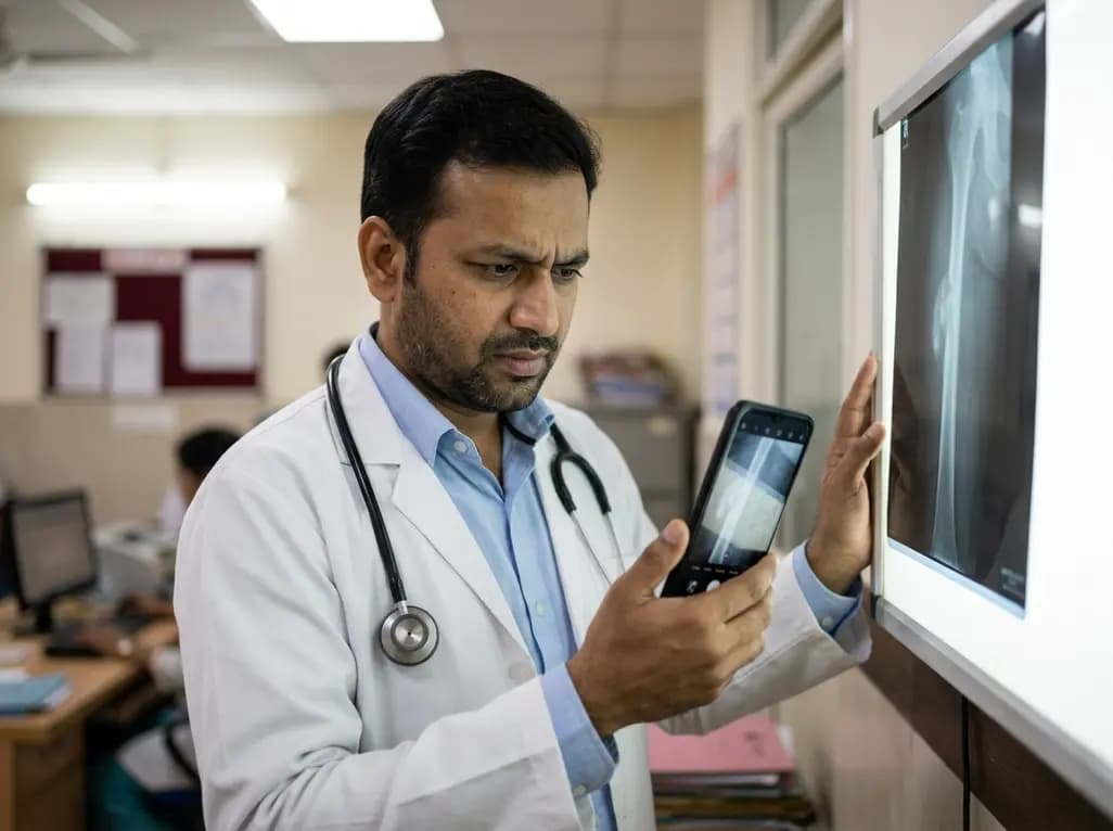
A retrospective study evaluating a locally deployed vision language model (Qwen3-VL) reveals that smartphone-captured radiographs and retrieval-augmented generation can paradoxically reduce diagnostic accuracy in bone tumor referrals due to optical noise and textual hallucinations.
Today
A real-world quasi-experimental study reveals that web-based apps fostering healthy habit formation significantly boost physical activity and dietary quality in adults. Discover key clinical insights on digital adherence, dose-response effects, and patient-specific modifiers in preventive medicine.
Today
