.webp&w=384&q=75)

Loading, please wait...
Loading, please wait...
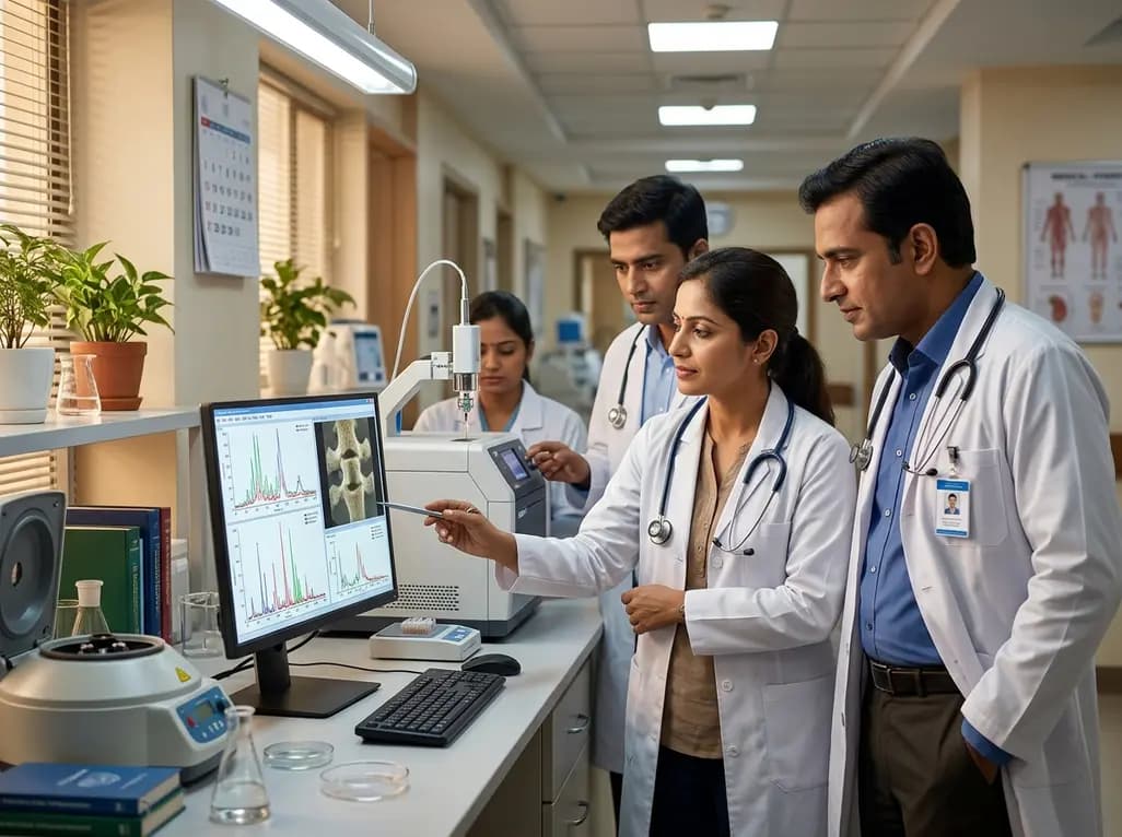
Surgical resection remains the primary therapeutic intervention for many extradural and intradural extramedullary spinal neoplasms. Achieving optimal patient outcomes requires precise intraoperative histopathological evaluation to guide the surgical extent. However, standard intraoperative frozen section analysis is time-consuming and inherently subjective. Emerging diagnostic innovations now offer rapid, objective molecular profiling. A landmark study published in Neuro-Oncology demonstrates that picosecond infrared laser mass spectrometry (PIRL-MS) delivers highly reliable spinal tumor diagnosis within just 10 seconds. This point-of-care tool provides actionable pathological classification directly in the operating room.
Intradural extramedullary spinal neoplasms constitute approximately 40% of all diagnosed spinal tumors, with schwannomas and meningiomas representing the vast majority. Clinicians face distinct surgical trade-offs depending on the exact histological subtype encountered. For instance, spinal meningiomas often benefit from aggressive resection including the dural attachment to prevent local recurrence. In contrast, schwannomas originate from nerve roots, where extensive resection risks unnecessary neurological morbidity. Therefore, surgeons rely heavily on rapid intraoperative consultations to calibrate their surgical margins.
Despite its widespread use, conventional frozen section analysis presents substantial logistical hurdles. The preparation and microscopic interpretation of frozen sections typically require 20 to 45 minutes, extending general anesthesia duration. Furthermore, tissue freezing artifacts can obscure critical cytoarchitectural details, leading to diagnostic ambiguity. Pathologist availability can also vary drastically, particularly during emergency or late-evening surgical procedures. These systemic limitations highlight an urgent demand for automated, objective diagnostic methods that assist surgical teams in real time without prolonged delays.
Picosecond infrared laser mass spectrometry leverages ultra-short laser pulses to vaporize microscopic volumes of tissue with negligible thermal damage. The laser selectively excites the vibrational modes of water molecules inside the cellular matrix, resulting in a gentle, instantaneous ablation termed desorption by ultrafast impulsive vibration. Consequently, intact biological molecules, including cellular lipids and metabolites, transfer directly into the gas phase without structural degradation.
Once transferred into the mass spectrometer, these biomolecules undergo ambient ionization and rapid spectral profiling. Within 10 seconds, the device captures a distinct mass spectrum that reflects the biochemical architecture of the sampled tissue. Unlike traditional diagnostic pipelines, PIRL-MS requires zero chemical fixation, staining, or laborious tissue processing. Because the laser extraction is so gentle, the fundamental lipid fingerprints remain intact, allowing computational algorithms to classify the specimen against pre-established spectral reference libraries almost instantaneously.
The core innovation behind PIRL-MS rests on the discovery of distinctive lipid signatures unique to each tumor class. In the study, researchers evaluated 319 clinical human specimens to build and validate a diagnostic classification model. High-resolution tandem mass spectrometry pinpointed a panel of 41 cellular lipids that serve as a molecular fingerprint for intraoperative classification.
This lipidomic panel predominantly encompasses four major structural classes: phosphatidylcholines, sphingomyelins, phosphatidylethanolamines, and ceramides. These structural lipids govern cell membrane fluidity, receptor clustering, and metabolic signaling pathways that differ between neoplastic lineages. Much like genomic, transcriptomic, or DNA-methylation arrays categorize central nervous system neoplasms, this lipid array provides robust diagnostic discrimination. However, while genomic profiling takes days, this lipid-based spinal tumor diagnosis delivers conclusive classification in seconds, democratizing molecular pathology at the surgical bedside.
In independent validation testing across 182 patient specimens, the PIRL-MS diagnostic model demonstrated exceptional performance. The platform achieved an overall diagnostic sensitivity of (93 ± 1)% and a specificity of (97 ± 2)% when distinguishing major spinal tumor types, including metastatic carcinomas, schwannomas, and meningiomas. These metrics match or exceed the diagnostic concordance typically reported for standard intraoperative frozen sections.
Moreover, the researchers rigorously tested the generalizability of the model against confounding pathologies. They introduced 54 additional intradural extramedullary spinal tumors, such as myxopapillary ependymomas, neurofibromas, paragangliomas, and solitary fibrous tumors. Remarkably, the accuracy of classifying meningiomas (n = 97) and schwannomas (n = 106) remained uncompromised despite the presence of previously unseen tumor types in the differential diagnosis. This robustness confirms that the 41-lipid biomarker array captures fundamental biological differences that resist distraction from rare histologic mimickers.
The ultra-rapid turnaround time of 10 seconds fundamentally alters intraoperative workflow and patient safety. Surgeons can sample multiple distinct zones within a tumor bed to verify margins without adding meaningful operative time. For spinal meningiomas, real-time confirmation empowers the surgical team to pursue radical resection and dural excision confidently. Conversely, confirming a schwannoma or non-malignant peripheral nerve sheath tumor allows the surgeon to preserve critical functional nerve fascicles.
Additionally, this objective technology helps mitigate diagnostic disparities in surgical centers that lack round-the-clock neuropathology subspecialists. Because the PIRL-MS system operates via automated classification algorithms, non-pathologists and surgical staff with basic laboratory training can perform the assay reliably. Consequently, community hospitals and resource-constrained centers could access tertiary-level diagnostic accuracy during complex spine surgeries, reducing reoperation rates and postoperative complications.
While the retrospective validation of PIRL-MS is compelling, ongoing research aims to integrate handheld laser probes directly into the sterile surgical field. Combining laser desorption mass spectrometry with real-time navigation systems could create an intelligent surgical scalpel capable of continuously sensing tissue margins. Furthermore, researchers are actively expanding the reference database to include rare pediatric spinal neoplasms and high-grade intramedullary lesions.
Prospective multicenter trials represent the next essential milestone before broad regulatory approval and commercial deployment. As machine learning algorithms continue to evolve, these dynamic molecular models will adapt to broader patient demographics and rare metabolic variants. Ultimately, ultrafast ambient mass spectrometry promises to bridge the gap between microscopic biochemistry and surgical precision, ushering in a new era of personalized intraoperative neuro-oncology.
PIRL-MS is an ultrafast analytical technology that uses picosecond laser pulses to ablate microscopic tissue samples without thermal injury. The vaporized molecules undergo instant mass spectrometry, generating a diagnostic biochemical profile in approximately 10 seconds without chemical stains or lengthy histological preparation.
Based on independent validation of human clinical specimens, PIRL-MS achieved a sensitivity of 93% and a specificity of 97%. It reliably differentiated common spinal tumor types, including meningiomas, schwannomas, and metastatic carcinomas, while maintaining high diagnostic stability against rare histological variants.
Yes. The platform utilizes automated machine learning algorithms to match lipid profiles against established diagnostic models. Consequently, surgical staff and technicians with basic laboratory training can operate the instrument, providing objective pathological insights in clinical settings with limited access to on-site neuropathologists.
Disclaimer: This content is for informational and educational purposes only and should not be used as a substitute for professional medical advice, diagnosis, or treatment. Always consult with a qualified healthcare provider regarding any medical condition or treatment options. Refer to the latest local and national guidelines for clinical practice.
References

Read summarized clinical updates, watch expert medical content, and earn CME certifications right from your smartphone.
Open your smartphone camera to scan and install.





A novel study demonstrates that picosecond infrared laser mass spectrometry (PIRL-MS) enables non-subjective spinal tumor diagnosis in 10 seconds. Using a 41-lipid molecular array, the technique achieved 93% sensitivity and 97% specificity for differentiating meningiomas, schwannomas, and metastatic carcinomas.
4 weeks back
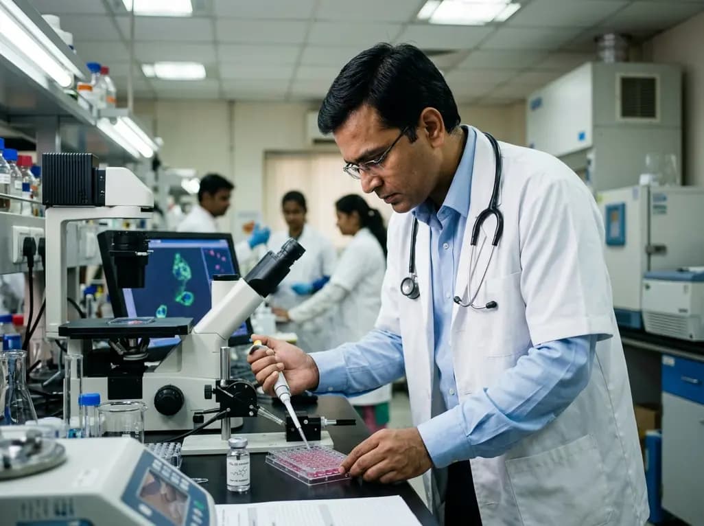
PZL-26, a selective small-molecule inhibitor of LONP1, reveals essential roles of this mitochondrial protease in complex I biogenesis, translation, and oxidative phosphorylation. These findings provide critical mechanistic insights for developing targeted anticancer therapies against proteostasis-dependent tumors.
Today

A nationwide South Korean study shows underreporting of smoking among women dropped from 57.4% to 36.4% between 2008 and 2021. However, over one-third of female smokers still conceal their habit, highlighting the critical role of cotinine verification in clinical risk assessment.
Today

PepsiCo and Monster Beverage have challenged the FSSAI ban on the energy drink label in Indian courts. While companies highlight supply disruptions, regulatory actions spotlight severe cardiovascular and metabolic risks associated with excessive caffeine, sugar, and taurine consumption, especially in adolescents.
Today
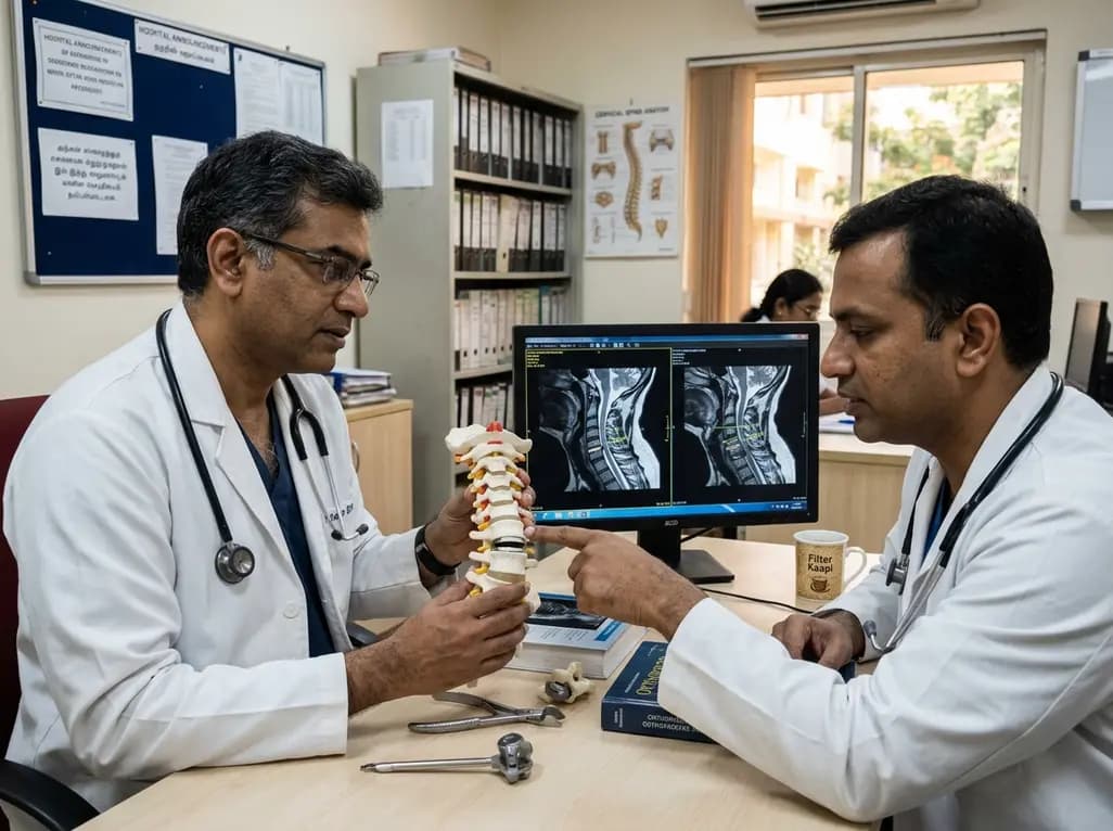
A recent investigation analyzes whether cervical disc arthroplasty effectively restores or merely preserves physiological range of motion, providing critical biomechanical insights for treating hypomobile cervical segments.
Today

A Bayesian multilevel meta-analysis reveals that aerobic training combined with moderate carbohydrate restriction modestly lowers HbA1c in type 2 diabetes. However, sparse data and very low certainty leave incremental benefits over exercise or diet alone unproven, highlighting the need for individualized care.
Yesterday
